 Movie
Movie Controller

[English] 日本語
 Yorodumi
- PDB-5yiw: Caulobacter crescentus GcrA DNA-binding domain (DBD) in complex w... -
+ Open data

- Basic information
| Entry | Database: PDB / ID: 5yiw | ||||||
|---|---|---|---|---|---|---|---|
| Title | Caulobacter crescentus GcrA DNA-binding domain (DBD) in complex with methylated dsDNA (crystal form 2) | ||||||
 Components Components |
| ||||||
 Keywords Keywords | DNA BINDING PROTEIN/DNA / Caulobacter crescentus / GcrA / DNA-binding / transcription factor / DNA BINDING PROTEIN / DNA BINDING PROTEIN-DNA complex | ||||||
| Function / homology | GcrA cell cycle regulator / GcrA cell cycle regulator / metal ion binding / (R,R)-2,3-BUTANEDIOL / DNA / DNA (> 10) / Cell cycle regulatory protein GcrA Function and homology information Function and homology information | ||||||
| Biological species |  Caulobacter crescentus (bacteria) Caulobacter crescentus (bacteria)synthetic construct (others) | ||||||
| Method |  X-RAY DIFFRACTION / SYNCHROTRON / MOLECULAR REPLACEMENT / Resolution: 1.551 Å X-RAY DIFFRACTION / SYNCHROTRON / MOLECULAR REPLACEMENT / Resolution: 1.551 Å | ||||||
 Authors Authors | Wu, X. / Zhang, Y. | ||||||
| Funding support |  China, 1items China, 1items
| ||||||
 Citation Citation | Journal: Nucleic Acids Res. / Year: 2018 Title: Structural insights into the unique mechanism of transcription activation by Caulobacter crescentus GcrA. Authors: Wu, X. / Haakonsen, D.L. / Sanderlin, A.G. / Liu, Y.J. / Shen, L. / Zhuang, N. / Laub, M.T. / Zhang, Y. | ||||||
| History |
|
- Structure visualization
| Structure viewer | Molecule: MolmilJmol/JSmol |
|---|
- Downloads & links
-Download
| PDBx/mmCIF format | 5yiw.cif.gz | 57.4 KB | Display | PDBx/mmCIF format |
|---|---|---|---|---|
| PDB format | pdb5yiw.ent.gz | 37.2 KB | Display | PDB format |
| PDBx/mmJSON format | 5yiw.json.gz | Tree view | PDBx/mmJSON format | |
| Others |  Other downloads Other downloads |
-Validation report
| Arichive directory | https://data.pdbj.org/pub/pdb/validation_reports/yi/5yiwftp://data.pdbj.org/pub/pdb/validation_reports/yi/5yiw | HTTPS FTP |
|---|
-Related structure data
| Related structure data |  5yiuSC  5yivC 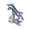 5yixC  5z7iC S: Starting model for refinement C: citing same article ( |
|---|---|
| Similar structure data |

-Links
 PDBj
PDBj


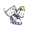
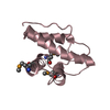
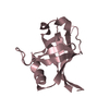
- Assembly
| Deposited unit | 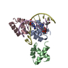
| ||||||||
|---|---|---|---|---|---|---|---|---|---|
| 1 | 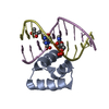
| ||||||||
| 2 | 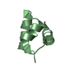
| ||||||||
| 3 | 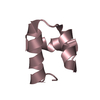
| ||||||||
| Unit cell |
|
-Components
| #1: Protein/peptide | Mass: 5317.199 Da / Num. of mol.: 3 / Fragment: DNA-binding domain (DBD) Source method: isolated from a genetically manipulated source Source: (gene. exp.) Caulobacter crescentus (strain NA1000 / CB15N) (bacteria)Strain: NA1000 / CB15N / Gene: gcrA, CCNA_02328 / Plasmid: pET28a / Production host: #2: DNA chain | | Mass: 3299.174 Da / Num. of mol.: 1 / Source method: obtained synthetically / Source: (synth.) synthetic construct (others) #3: DNA chain | | Mass: 3397.250 Da / Num. of mol.: 1 / Source method: obtained synthetically / Source: (synth.) synthetic construct (others) #4: Chemical | ChemComp-BU3 / ( |   Mass: 90.121 Da / Num. of mol.: 1 / Source method: obtained synthetically / Formula: C4H10O2 Mass: 90.121 Da / Num. of mol.: 1 / Source method: obtained synthetically / Formula: C4H10O2#5: Water | ChemComp-HOH / |  Mass: 18.015 Da / Num. of mol.: 119 / Source method: isolated from a natural source / Formula: H2O Mass: 18.015 Da / Num. of mol.: 119 / Source method: isolated from a natural source / Formula: H2O |
|---|
-Experimental details
-Experiment
| Experiment | Method: X-RAY DIFFRACTION / Number of used crystals: 1 |
|---|
- Sample preparation
| Crystal | Density Matthews: 2.21 Å3/Da / Density % sol: 44.36 % |
|---|---|
| Crystal grow | Temperature: 295 K / Method: vapor diffusion, hanging drop / Details: 20% PEG 8000, 0.05M Potassium phosphate monobasic |
-Data collection
| Diffraction | Mean temperature: 100 K |
|---|---|
| Diffraction source | Source: SYNCHROTRON / Site: SSRF / Beamline: BL17U1 / Wavelength: 0.979 Å |
| Detector | Type: DECTRIS PILATUS3 S 6M / Detector: PIXEL / Date: May 8, 2016 |
| Radiation | Protocol: SINGLE WAVELENGTH / Monochromatic (M) / Laue (L): M / Scattering type: x-ray |
| Radiation wavelength | Wavelength: 0.979 Å / Relative weight: 1 |
| Reflection | Resolution: 1.55→50 Å / Num. obs: 29530 / % possible obs: 99.6 % / Redundancy: 7.2 % / Biso Wilson estimate: 19.46 Å2 / Rmerge(I) obs: 0.07 / Rpim(I) all: 0.03 / Rrim(I) all: 0.07 / Rsym value: 0.07 / Χ2: 0.95 / Net I/σ(I): 22.5 |
| Reflection shell | Resolution: 1.55→1.58 Å / Redundancy: 6.9 % / Rmerge(I) obs: 0.74 / Mean I/σ(I) obs: 2.5 / Num. unique obs: 1394 / CC1/2: 0.88 / Rpim(I) all: 0.3 / Rrim(I) all: 0.8 / Rsym value: 0.74 / Χ2: 0.95 / % possible all: 99.9 |
- Processing
| Software |
| ||||||||||||||||||||||||||||||||||||||||||||||||||||||||||||||||||||||||||||||||||||
|---|---|---|---|---|---|---|---|---|---|---|---|---|---|---|---|---|---|---|---|---|---|---|---|---|---|---|---|---|---|---|---|---|---|---|---|---|---|---|---|---|---|---|---|---|---|---|---|---|---|---|---|---|---|---|---|---|---|---|---|---|---|---|---|---|---|---|---|---|---|---|---|---|---|---|---|---|---|---|---|---|---|---|---|---|---|
| Refinement | Method to determine structure: MOLECULAR REPLACEMENT Starting model: 5YIU Resolution: 1.551→28.745 Å / SU ML: 0.15 / Cross valid method: FREE R-VALUE / σ(F): 1.35 / Phase error: 25.68
| ||||||||||||||||||||||||||||||||||||||||||||||||||||||||||||||||||||||||||||||||||||
| Solvent computation | Shrinkage radii: 0.9 Å / VDW probe radii: 1.11 Å | ||||||||||||||||||||||||||||||||||||||||||||||||||||||||||||||||||||||||||||||||||||
| Displacement parameters | Biso mean: 27.7 Å2 | ||||||||||||||||||||||||||||||||||||||||||||||||||||||||||||||||||||||||||||||||||||
| Refinement step | Cycle: LAST / Resolution: 1.551→28.745 Å
| ||||||||||||||||||||||||||||||||||||||||||||||||||||||||||||||||||||||||||||||||||||
| Refine LS restraints |
| ||||||||||||||||||||||||||||||||||||||||||||||||||||||||||||||||||||||||||||||||||||
| LS refinement shell |
|
