| Entry | Database: PDB / ID: 5yh8
|
|---|
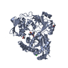
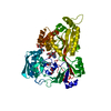
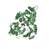
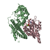
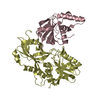
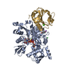
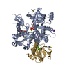
| Title | The crystal structure of Staphylococcus aureus CntA in complex with staphylopine and nickel |
|---|
 Components Components | Nickel ABC transporter substrate-binding protein |
|---|
 Keywords Keywords | METAL BINDING PROTEIN / complex / receptor |
|---|
| Function / homology |  Function and homology information Function and homology information
nickel cation transport / cobalt ion transport / zinc ion transport / peptide transport / peptide transmembrane transporter activity / nickel cation binding / ATP-binding cassette (ABC) transporter complex / outer membrane-bounded periplasmic space / heme bindingSimilarity search - Function Nickel ABC transporter, substrate-binding protein NikA / Solute-binding protein family 5, conserved site / Bacterial extracellular solute-binding proteins, family 5 signature. / Peptide/nickel binding protein, MppA-type / Solute-binding protein family 5 domain / Solute-binding protein family 5 / Bacterial extracellular solute-binding proteins, family 5 Middle / Periplasmic binding protein-like II / D-Maltodextrin-Binding Protein; domain 2 / Prokaryotic membrane lipoprotein lipid attachment site profile. ...Nickel ABC transporter, substrate-binding protein NikA / Solute-binding protein family 5, conserved site / Bacterial extracellular solute-binding proteins, family 5 signature. / Peptide/nickel binding protein, MppA-type / Solute-binding protein family 5 domain / Solute-binding protein family 5 / Bacterial extracellular solute-binding proteins, family 5 Middle / Periplasmic binding protein-like II / D-Maltodextrin-Binding Protein; domain 2 / Prokaryotic membrane lipoprotein lipid attachment site profile. / 3-Layer(aba) Sandwich / Alpha BetaSimilarity search - Domain/homology Chem-8UX / NICKEL (II) ION / DI(HYDROXYETHYL)ETHER / Nickel ABC transporter substrate-binding protein / Metal-staphylopine-binding protein CntASimilarity search - Component |
|---|
| Biological species |   Staphylococcus aureus (bacteria) Staphylococcus aureus (bacteria) |
|---|
| Method |  X-RAY DIFFRACTION / SYNCHROTRON / MOLECULAR REPLACEMENT / Resolution: 2.12 Å X-RAY DIFFRACTION / SYNCHROTRON / MOLECULAR REPLACEMENT / Resolution: 2.12 Å |
|---|
 Authors Authors | Ji, Q. / Song, L. |
|---|
 Citation Citation | Journal: Proc. Natl. Acad. Sci. U.S.A. / Year: 2018
Title: Mechanistic insights into staphylopine-mediated metal acquisition
Authors: Song, L. / Zhang, Y. / Chen, W. / Gu, T. / Zhang, S.Y. / Ji, Q. |
|---|
| History | | Deposition | Sep 27, 2017 | Deposition site: PDBJ / Processing site: PDBJ |
|---|
| Revision 1.0 | Mar 28, 2018 | Provider: repository / Type: Initial release |
|---|
| Revision 1.1 | Apr 11, 2018 | Group: Data collection / Database references / Category: citation
Item: _citation.journal_abbrev / _citation.pdbx_database_id_PubMed |
|---|
| Revision 1.2 | Apr 25, 2018 | Group: Data collection / Database references / Category: citation
Item: _citation.journal_volume / _citation.page_first / _citation.page_last |
|---|
| Revision 1.3 | Mar 27, 2024 | Group: Data collection / Database references / Category: chem_comp_atom / chem_comp_bond / database_2
Item: _database_2.pdbx_DOI / _database_2.pdbx_database_accession |
|---|
|
|---|
 Movie
Movie Controller

 Yorodumi
 Open data
 Basic information
 Structure visualization
 Downloads & links
 Other downloads


 PDBj
PDBj


 Assembly
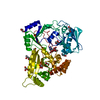


 Mass: 58.693 Da / Num. of mol.: 1 / Source method: obtained synthetically / Formula: Ni
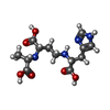
 Mass: 328.321 Da / Num. of mol.: 1 / Source method: obtained synthetically / Formula: C13H20N4O6
 Mass: 92.094 Da / Num. of mol.: 9 / Source method: obtained synthetically / Formula: C3H8O3
 Mass: 106.120 Da / Num. of mol.: 2 / Source method: obtained synthetically / Formula: C4H10O3
 Sample preparation
 / Beamline: BL19U1 / Wavelength: 0.9785 Å
 Processing