 Movie
Movie Controller

[English] 日本語
 Yorodumi
- PDB-5yg9: Crystal structure of ribose-1,5-bisphosphate isomerase mutant C13... -
+ Open data

- Basic information
| Entry | Database: PDB / ID: 5yg9 | |||||||||
|---|---|---|---|---|---|---|---|---|---|---|
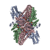
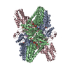
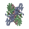
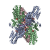
| Title | Crystal structure of ribose-1,5-bisphosphate isomerase mutant C135S from Pyrococcus horikoshii OT3 in complex with ribose-1,5-bisphosphate, AMP and GMP | |||||||||
 Components Components | Ribose 1,5-bisphosphate isomerase | |||||||||
 Keywords Keywords | ISOMERASE / NMP degradation pathway / Ribose-1 / 5-bisphosphate / Rossmann-like fold | |||||||||
| Function / homology |  Function and homology information Function and homology informationribose-1,5-bisphosphate isomerase / ribose 1,5-bisphosphate isomerase activity / S-methyl-5-thioribose-1-phosphate isomerase activity / pentose catabolic process / : Similarity search - Function | |||||||||
| Biological species |   Pyrococcus horikoshii (archaea) Pyrococcus horikoshii (archaea) | |||||||||
| Method |  X-RAY DIFFRACTION / MOLECULAR REPLACEMENT / molecular replacement / Resolution: 2.8 Å X-RAY DIFFRACTION / MOLECULAR REPLACEMENT / molecular replacement / Resolution: 2.8 Å | |||||||||
 Authors Authors | Gogoi, P. / Kanaujia, S.P. | |||||||||
| Funding support |  India, 1items India, 1items
| |||||||||
 Citation Citation | Journal: Sci Rep / Year: 2018 Title: A presumed homologue of the regulatory subunits of eIF2B functions as ribose-1,5-bisphosphate isomerase in Pyrococcus horikoshii OT3. Authors: Gogoi, P. / Kanaujia, S.P. | |||||||||
| History |
|
- Structure visualization
| Structure viewer | Molecule: MolmilJmol/JSmol |
|---|
- Downloads & links
-Download
| PDBx/mmCIF format | 5yg9.cif.gz | 207.2 KB | Display | PDBx/mmCIF format |
|---|---|---|---|---|
| PDB format | pdb5yg9.ent.gz | 165.2 KB | Display | PDB format |
| PDBx/mmJSON format | 5yg9.json.gz | Tree view | PDBx/mmJSON format | |
| Others |  Other downloads Other downloads |
-Validation report
| Arichive directory | https://data.pdbj.org/pub/pdb/validation_reports/yg/5yg9ftp://data.pdbj.org/pub/pdb/validation_reports/yg/5yg9 | HTTPS FTP |
|---|
-Related structure data
| Related structure data |  5yfjSC 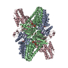 5yfsC  5yftC 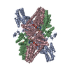 5yfuC  5yfvC  5yfwC 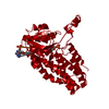 5yfxC 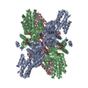 5yg5C  5yg6C  5yg7C  5yg8C  5ygaC S: Starting model for refinement C: citing same article ( |
|---|---|
| Similar structure data |


-Links
 PDBj
PDBj


- Assembly
| Deposited unit | 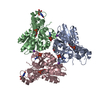
| ||||||||
|---|---|---|---|---|---|---|---|---|---|
| 1 | 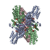
| ||||||||
| Unit cell |
|
-Components
-Protein / Sugars , 2 types, 6 molecules ABC
| #1: Protein | Mass: 36440.289 Da / Num. of mol.: 3 / Mutation: C135S Source method: isolated from a genetically manipulated source Source: (gene. exp.) Pyrococcus horikoshii (archaea)Strain: ATCC 700860 / DSM 12428 / JCM 9974 / NBRC 100139 / OT-3 Gene: PH0208 / Plasmid: pET22b / Production host:  References: UniProt: O57947, ribose-1,5-bisphosphate isomerase #2: Sugar |  Type: D-saccharide, alpha linking / Mass: 310.090 Da / Num. of mol.: 3 / Source method: obtained synthetically / Formula: C5H12O11P2 / Feature type: SUBJECT OF INVESTIGATION Type: D-saccharide, alpha linking / Mass: 310.090 Da / Num. of mol.: 3 / Source method: obtained synthetically / Formula: C5H12O11P2 / Feature type: SUBJECT OF INVESTIGATION |
|---|
-Non-polymers , 5 types, 219 molecules 


| #3: Chemical |  Mass: 347.221 Da / Num. of mol.: 3 / Source method: obtained synthetically / Formula: C10H14N5O7P / Feature type: SUBJECT OF INVESTIGATION / Comment: AMP*YM Mass: 347.221 Da / Num. of mol.: 3 / Source method: obtained synthetically / Formula: C10H14N5O7P / Feature type: SUBJECT OF INVESTIGATION / Comment: AMP*YM#4: Chemical |  Mass: 39.098 Da / Num. of mol.: 3 / Source method: obtained synthetically / Formula: K Mass: 39.098 Da / Num. of mol.: 3 / Source method: obtained synthetically / Formula: K#5: Chemical | ChemComp-MPD / ( |  Mass: 118.174 Da / Num. of mol.: 1 / Source method: obtained synthetically / Formula: C6H14O2 / Comment: precipitant*YM Mass: 118.174 Da / Num. of mol.: 1 / Source method: obtained synthetically / Formula: C6H14O2 / Comment: precipitant*YM#6: Chemical |  Mass: 363.221 Da / Num. of mol.: 2 / Source method: obtained synthetically / Formula: C10H14N5O8P / Feature type: SUBJECT OF INVESTIGATION Mass: 363.221 Da / Num. of mol.: 2 / Source method: obtained synthetically / Formula: C10H14N5O8P / Feature type: SUBJECT OF INVESTIGATION#7: Water | ChemComp-HOH / | Mass: 18.015 Da / Num. of mol.: 210 / Source method: isolated from a natural source / Formula: H2O |
|---|
-Experimental details
-Experiment
| Experiment | Method: X-RAY DIFFRACTION / Number of used crystals: 1 |
|---|
- Sample preparation
| Crystal | Density Matthews: 3.31 Å3/Da / Density % sol: 62.85 % |
|---|---|
| Crystal grow | Temperature: 293 K / Method: vapor diffusion, hanging drop / Details: 0.7 M NaCl, 3% PEG 6000, 30% MPD |
-Data collection
| Diffraction | Mean temperature: 100 K | ||||||||||||||||||||||||
|---|---|---|---|---|---|---|---|---|---|---|---|---|---|---|---|---|---|---|---|---|---|---|---|---|---|
| Diffraction source | Source: ROTATING ANODE / Type: RIGAKU MICROMAX-007 HF / Wavelength: 1.5418 Å | ||||||||||||||||||||||||
| Detector | Type: RIGAKU RAXIS IV++ / Detector: IMAGE PLATE / Date: Mar 10, 2017 / Details: VariMax HF | ||||||||||||||||||||||||
| Radiation | Protocol: SINGLE WAVELENGTH / Monochromatic (M) / Laue (L): M / Scattering type: x-ray | ||||||||||||||||||||||||
| Radiation wavelength | Wavelength: 1.5418 Å / Relative weight: 1 | ||||||||||||||||||||||||
| Reflection | Resolution: 2.8→85.6 Å / Num. obs: 35799 / % possible obs: 100 % / Redundancy: 11.7 % / CC1/2: 0.997 / Rmerge(I) obs: 0.147 / Rpim(I) all: 0.045 / Rrim(I) all: 0.154 / Net I/σ(I): 15.6 | ||||||||||||||||||||||||
| Reflection shell | Diffraction-ID: 1 / % possible all: 100
|
-Phasing
| Phasing | Method: molecular replacement | |||||||||
|---|---|---|---|---|---|---|---|---|---|---|
| Phasing MR | Model details: Phaser MODE: MR_AUTO
|
- Processing
| Software |
| ||||||||||||||||||||||||||||||||||||||||||||||||||||||||||||
|---|---|---|---|---|---|---|---|---|---|---|---|---|---|---|---|---|---|---|---|---|---|---|---|---|---|---|---|---|---|---|---|---|---|---|---|---|---|---|---|---|---|---|---|---|---|---|---|---|---|---|---|---|---|---|---|---|---|---|---|---|---|
| Refinement | Method to determine structure: MOLECULAR REPLACEMENT Starting model: 5YFJ Resolution: 2.8→85.6 Å / Cor.coef. Fo:Fc: 0.946 / Cor.coef. Fo:Fc free: 0.897 / SU B: 11.309 / SU ML: 0.221 / SU R Cruickshank DPI: 0.915 / Cross valid method: THROUGHOUT / σ(F): 0 / ESU R: 0.915 / ESU R Free: 0.314 Details: HYDROGENS HAVE BEEN ADDED IN THE RIDING POSITIONS U VALUES : REFINED INDIVIDUALLY
| ||||||||||||||||||||||||||||||||||||||||||||||||||||||||||||
| Solvent computation | Ion probe radii: 0.8 Å / Shrinkage radii: 0.8 Å / VDW probe radii: 1.2 Å | ||||||||||||||||||||||||||||||||||||||||||||||||||||||||||||
| Displacement parameters | Biso max: 148.79 Å2 / Biso mean: 37.725 Å2 / Biso min: 6.69 Å2
| ||||||||||||||||||||||||||||||||||||||||||||||||||||||||||||
| Refinement step | Cycle: final / Resolution: 2.8→85.6 Å
| ||||||||||||||||||||||||||||||||||||||||||||||||||||||||||||
| Refine LS restraints |
| ||||||||||||||||||||||||||||||||||||||||||||||||||||||||||||
| LS refinement shell | Resolution: 2.8→2.873 Å / Rfactor Rfree error: 0 / Total num. of bins used: 20
|
