 Movie
Movie Controller


+ Open data

- Basic information
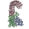
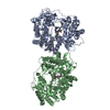
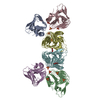
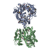
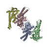
| Entry | Database: PDB / ID: 5yet | ||||||
|---|---|---|---|---|---|---|---|
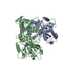
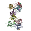
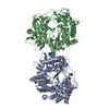
| Title | Structure of R354_WT | ||||||
 Components Components | Uncharacterized protein R354 | ||||||
 Keywords Keywords | NUCLEAR PROTEIN / MIMIVIRE / Cas4-like / nuclease / R354 | ||||||
| Function / homology | Putative phage-type endonuclease / YqaJ viral recombinase / : / YqaJ-like viral recombinase domain / PD-(D/E)XK endonuclease-like domain superfamily / Restriction endonuclease type II-like / Uncharacterized protein R354 Function and homology information Function and homology information | ||||||
| Biological species |  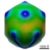 Acanthamoeba polyphaga mimivirus Acanthamoeba polyphaga mimivirus | ||||||
| Method |  X-RAY DIFFRACTION / SYNCHROTRON / SAD / Resolution: 2.806 Å X-RAY DIFFRACTION / SYNCHROTRON / SAD / Resolution: 2.806 Å | ||||||
 Authors Authors | Dou, C. / Yu, M.J. / Gu, Y.J. / Cheng, W. | ||||||
 Citation Citation | Journal: Iscience / Year: 2018 Title: Structural and Mechanistic Analyses Reveal a Unique Cas4-like Protein in the Mimivirus Virophage Resistance Element System. Authors: Dou, C. / Yu, M. / Gu, Y. / Wang, J. / Yin, K. / Nie, C. / Zhu, X. / Qi, S. / Wei, Y. / Cheng, W. | ||||||
| History |
|
- Structure visualization
| Structure viewer | Molecule: MolmilJmol/JSmol |
|---|
- Downloads & links
-Download
| PDBx/mmCIF format | 5yet.cif.gz | 172.2 KB | Display | PDBx/mmCIF format |
|---|---|---|---|---|
| PDB format | pdb5yet.ent.gz | 137.3 KB | Display | PDB format |
| PDBx/mmJSON format | 5yet.json.gz | Tree view | PDBx/mmJSON format | |
| Others |  Other downloads Other downloads |
-Validation report
| Arichive directory | https://data.pdbj.org/pub/pdb/validation_reports/ye/5yetftp://data.pdbj.org/pub/pdb/validation_reports/ye/5yet | HTTPS FTP |
|---|
-Related structure data
-Links
 PDBj
PDBj


- Assembly
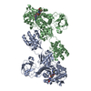
| Deposited unit | 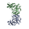
| ||||||||
|---|---|---|---|---|---|---|---|---|---|
| 1 |
| ||||||||
| Unit cell |
|
-Components
| #1: Protein | Mass: 47234.547 Da / Num. of mol.: 2 / Fragment: UNP RESIDUES 139-537 Source method: isolated from a genetically manipulated source Source: (gene. exp.) Acanthamoeba polyphaga mimivirus / Gene: MIMI_R354 / Production host:  #2: Water | ChemComp-HOH / |  Mass: 18.015 Da / Num. of mol.: 49 / Source method: isolated from a natural source / Formula: H2O Mass: 18.015 Da / Num. of mol.: 49 / Source method: isolated from a natural source / Formula: H2O |
|---|
-Experimental details
-Experiment
| Experiment | Method: X-RAY DIFFRACTION / Number of used crystals: 1 |
|---|
- Sample preparation
| Crystal | Density Matthews: 2.86 Å3/Da / Density % sol: 56.92 % |
|---|---|
| Crystal grow | Temperature: 293 K / Method: vapor diffusion, sitting drop / Details: 200mM ammonium tartrate, 20% PEG 3350 |
-Data collection
| Diffraction | Mean temperature: 100 K |
|---|---|
| Diffraction source | Source: SYNCHROTRON / Site: SSRF  / Beamline: BL19U1 / Wavelength: 0.9785 Å / Beamline: BL19U1 / Wavelength: 0.9785 Å |
| Detector | Type: DECTRIS PILATUS3 S 6M / Detector: PIXEL / Date: Jun 12, 2016 |
| Radiation | Protocol: SINGLE WAVELENGTH / Monochromatic (M) / Laue (L): M / Scattering type: x-ray |
| Radiation wavelength | Wavelength: 0.9785 Å / Relative weight: 1 |
| Reflection | Resolution: 2.806→48.949 Å / Num. obs: 22531 / % possible obs: 85.48 % / Redundancy: 4.8 % / Net I/σ(I): 13.05 |
- Processing
| Software |
| |||||||||||||||||||||||||||||||||||||||||||||||||||||||||||||||
|---|---|---|---|---|---|---|---|---|---|---|---|---|---|---|---|---|---|---|---|---|---|---|---|---|---|---|---|---|---|---|---|---|---|---|---|---|---|---|---|---|---|---|---|---|---|---|---|---|---|---|---|---|---|---|---|---|---|---|---|---|---|---|---|---|
| Refinement | Method to determine structure: SAD / Resolution: 2.806→48.949 Å / SU ML: 0.34 / Cross valid method: FREE R-VALUE / σ(F): 1.35 / Phase error: 24.93
| |||||||||||||||||||||||||||||||||||||||||||||||||||||||||||||||
| Solvent computation | Shrinkage radii: 0.9 Å / VDW probe radii: 1.11 Å | |||||||||||||||||||||||||||||||||||||||||||||||||||||||||||||||
| Refinement step | Cycle: LAST / Resolution: 2.806→48.949 Å
| |||||||||||||||||||||||||||||||||||||||||||||||||||||||||||||||
| Refine LS restraints |
| |||||||||||||||||||||||||||||||||||||||||||||||||||||||||||||||
| LS refinement shell |
|
