 Movie
Movie Controller

[English] 日本語
 Yorodumi
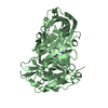
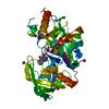
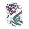
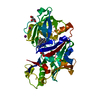
- PDB-5ycz: Crystal structure of Alocasin, protease inhibitor from Giant Taro... -
+ Open data

- Basic information
| Entry | Database: PDB / ID: 5ycz | ||||||
|---|---|---|---|---|---|---|---|
| Title | Crystal structure of Alocasin, protease inhibitor from Giant Taro (Arum macrorrhizon) | ||||||
 Components Components | Trypsin/chymotrypsin inhibitor | ||||||
 Keywords Keywords | PLANT PROTEIN / Protease inhibitor / Storage protein / Tubers / Alocasia / Alocasin / Kunitz type | ||||||
| Function / homology | Proteinase inhibitor A/B / Proteinase inhibitor I3, Kunitz legume / Trypsin and protease inhibitor / Soybean trypsin inhibitor (Kunitz) family of protease inhibitors / Kunitz inhibitor STI-like superfamily / serine-type endopeptidase inhibitor activity / Trypsin/chymotrypsin inhibitor Function and homology information Function and homology information | ||||||
| Biological species |  Alocasia macrorrhizos (giant taro) Alocasia macrorrhizos (giant taro) | ||||||
| Method |  X-RAY DIFFRACTION / SYNCHROTRON / MOLECULAR REPLACEMENT / Resolution: 2.502 Å X-RAY DIFFRACTION / SYNCHROTRON / MOLECULAR REPLACEMENT / Resolution: 2.502 Å | ||||||
 Authors Authors | Vajravijayan, S. / Pletnev, S. / Nandhagopal, N. / Gunasekaran, K. | ||||||
 Citation Citation | Journal: Pest Manag. Sci. / Year: 2018 Title: Crystal structure of a novel Kunitz type inhibitor, alocasin with anti-Aedes aegypti activity targeting midgut proteases. Authors: Vajravijayan, S. / Pletnev, S. / Pletnev, V.Z. / Nandhagopal, N. / Gunasekaran, K. | ||||||
| History |
|
- Structure visualization
| Structure viewer | Molecule: MolmilJmol/JSmol |
|---|
- Downloads & links
-Download
| PDBx/mmCIF format | 5ycz.cif.gz | 80.1 KB | Display | PDBx/mmCIF format |
|---|---|---|---|---|
| PDB format | pdb5ycz.ent.gz | 60.5 KB | Display | PDB format |
| PDBx/mmJSON format | 5ycz.json.gz | Tree view | PDBx/mmJSON format | |
| Others |  Other downloads Other downloads |
-Validation report
| Arichive directory | https://data.pdbj.org/pub/pdb/validation_reports/yc/5yczftp://data.pdbj.org/pub/pdb/validation_reports/yc/5ycz | HTTPS FTP |
|---|
-Related structure data
| Similar structure data |
|---|


-Links
 PDBj
PDBj- Assembly
| Deposited unit | 
| ||||||||
|---|---|---|---|---|---|---|---|---|---|
| 1 |
| ||||||||
| Unit cell |
|
-Components
| #1: Protein | Mass: 19793.139 Da / Num. of mol.: 2 / Source method: isolated from a natural source / Source: (natural) Alocasia macrorrhizos (giant taro) / References: UniProt: P35812#2: Water | ChemComp-HOH / |  Mass: 18.015 Da / Num. of mol.: 102 / Source method: isolated from a natural source / Formula: H2O Mass: 18.015 Da / Num. of mol.: 102 / Source method: isolated from a natural source / Formula: H2OHas protein modification | Y | |
|---|
-Experimental details
-Experiment
| Experiment | Method: X-RAY DIFFRACTION / Number of used crystals: 1 |
|---|
- Sample preparation
| Crystal | Density Matthews: 2.49 Å3/Da / Density % sol: 50.64 % |
|---|---|
| Crystal grow | Temperature: 293 K / Method: vapor diffusion, hanging drop / pH: 7.5 / Details: 0.1M HEPES (pH7.5). 1.6 M Ammonium Sulfate |
-Data collection
| Diffraction | Mean temperature: 100 K | |||||||||||||||||||||||||||||||||||||||||||||||||||||||||||||||||||||||||||||||||||||||||||||||||||
|---|---|---|---|---|---|---|---|---|---|---|---|---|---|---|---|---|---|---|---|---|---|---|---|---|---|---|---|---|---|---|---|---|---|---|---|---|---|---|---|---|---|---|---|---|---|---|---|---|---|---|---|---|---|---|---|---|---|---|---|---|---|---|---|---|---|---|---|---|---|---|---|---|---|---|---|---|---|---|---|---|---|---|---|---|---|---|---|---|---|---|---|---|---|---|---|---|---|---|---|---|
| Diffraction source | Source: SYNCHROTRON / Site: APS  / Beamline: 22-ID / Wavelength: 1 Å / Beamline: 22-ID / Wavelength: 1 Å | |||||||||||||||||||||||||||||||||||||||||||||||||||||||||||||||||||||||||||||||||||||||||||||||||||
| Detector | Type: MARMOSAIC 300 mm CCD / Detector: CCD / Date: Mar 7, 2017 | |||||||||||||||||||||||||||||||||||||||||||||||||||||||||||||||||||||||||||||||||||||||||||||||||||
| Radiation | Protocol: SINGLE WAVELENGTH / Monochromatic (M) / Laue (L): M / Scattering type: x-ray | |||||||||||||||||||||||||||||||||||||||||||||||||||||||||||||||||||||||||||||||||||||||||||||||||||
| Radiation wavelength | Wavelength: 1 Å / Relative weight: 1 | |||||||||||||||||||||||||||||||||||||||||||||||||||||||||||||||||||||||||||||||||||||||||||||||||||
| Reflection | Resolution: 2.5→30 Å / Num. obs: 14759 / % possible obs: 99.2 % / Redundancy: 5.7 % / Rmerge(I) obs: 0.095 / Rpim(I) all: 0.042 / Rrim(I) all: 0.104 / Χ2: 0.89 / Net I/σ(I): 8.4 / Num. measured all: 83766 | |||||||||||||||||||||||||||||||||||||||||||||||||||||||||||||||||||||||||||||||||||||||||||||||||||
| Reflection shell | Diffraction-ID: 1
|
- Processing
| Software |
| ||||||||||||||||||||||||
|---|---|---|---|---|---|---|---|---|---|---|---|---|---|---|---|---|---|---|---|---|---|---|---|---|---|
| Refinement | Method to determine structure: MOLECULAR REPLACEMENT / Resolution: 2.502→29.354 Å / SU ML: 0.34 / Cross valid method: THROUGHOUT / σ(F): 1.35 / Phase error: 28.6 / Stereochemistry target values: ML
| ||||||||||||||||||||||||
| Solvent computation | Shrinkage radii: 0.9 Å / VDW probe radii: 1.11 Å / Solvent model: FLAT BULK SOLVENT MODEL | ||||||||||||||||||||||||
| Displacement parameters | Biso max: 93.12 Å2 / Biso mean: 41.3613 Å2 / Biso min: 13.5 Å2 | ||||||||||||||||||||||||
| Refinement step | Cycle: final / Resolution: 2.502→29.354 Å
|
