 Movie
Movie Controller


+ Open data

- Basic information
| Entry | Database: PDB / ID: 5y8f | ||||||
|---|---|---|---|---|---|---|---|
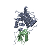
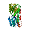
| Title | Crystal Structure of a prokaryotic SEFIR domain | ||||||
 Components Components | Sefir domain protein | ||||||
 Keywords Keywords | SIGNALING PROTEIN / SEFIR | ||||||
| Function / homology | SEFIR domain / SEFIR domain profile. / NAD+ catabolic process / NAD+ nucleosidase activity / TIR domain / Toll/interleukin-1 receptor homology (TIR) domain / Toll/interleukin-1 receptor homology (TIR) domain superfamily / signal transduction / Sefir domain protein Function and homology information Function and homology information | ||||||
| Biological species |  | ||||||
| Method |  X-RAY DIFFRACTION / MOLECULAR REPLACEMENT / Resolution: 2 Å X-RAY DIFFRACTION / MOLECULAR REPLACEMENT / Resolution: 2 Å | ||||||
 Authors Authors | Zhang, R. / Ye, S. / Zhu, Y. / Yang, H. | ||||||
 Citation Citation | Journal: J. Struct. Biol. / Year: 2018 Title: Structure of a prokaryotic SEFIR domain reveals two novel SEFIR-SEFIR interaction modes. Authors: Yang, H. / Zhu, Y. / Chen, X. / Li, X. / Ye, S. / Zhang, R. | ||||||
| History |
|
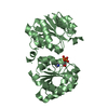
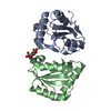
- Structure visualization
| Structure viewer | Molecule: MolmilJmol/JSmol |
|---|
- Downloads & links
-Download
| PDBx/mmCIF format | 5y8f.cif.gz | 79.6 KB | Display | PDBx/mmCIF format |
|---|---|---|---|---|
| PDB format | pdb5y8f.ent.gz | 58.9 KB | Display | PDB format |
| PDBx/mmJSON format | 5y8f.json.gz | Tree view | PDBx/mmJSON format | |
| Others |  Other downloads Other downloads |
-Validation report
| Summary document | 5y8f_validation.pdf.gz | 435 KB | Display | wwPDB validaton report |
|---|---|---|---|---|
| Full document | 5y8f_full_validation.pdf.gz | 438.7 KB | Display | |
| Data in XML | 5y8f_validation.xml.gz | 16.7 KB | Display | |
| Data in CIF | 5y8f_validation.cif.gz | 24.6 KB | Display | |
| Arichive directory | https://data.pdbj.org/pub/pdb/validation_reports/y8/5y8fftp://data.pdbj.org/pub/pdb/validation_reports/y8/5y8f | HTTPS FTP |
-Related structure data


-Links
 PDBj
PDBj
- Assembly
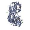
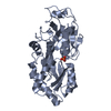
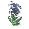
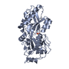
| Deposited unit | 
| ||||||||
|---|---|---|---|---|---|---|---|---|---|
| 1 |
| ||||||||
| Unit cell |
|
-Components
| #1: Protein | Mass: 16953.119 Da / Num. of mol.: 2 / Fragment: UNP RESIDUES 8-150 Source method: isolated from a genetically manipulated source Source: (gene. exp.) #2: Water | ChemComp-HOH / |  Mass: 18.015 Da / Num. of mol.: 346 / Source method: isolated from a natural source / Formula: H2O Mass: 18.015 Da / Num. of mol.: 346 / Source method: isolated from a natural source / Formula: H2O |
|---|
-Experimental details
-Experiment
| Experiment | Method: X-RAY DIFFRACTION / Number of used crystals: 1 |
|---|
- Sample preparation
| Crystal | Density Matthews: 2.07 Å3/Da / Density % sol: 40.53 % |
|---|---|
| Crystal grow | Temperature: 289 K / Method: vapor diffusion, hanging drop / Details: 0.2 M NaCl, 0.1 M imidozale pH 8.0, 30% PEG 8000 |
-Data collection
| Diffraction | Mean temperature: 173 K |
|---|---|
| Diffraction source | Source: ROTATING ANODE / Type: RIGAKU FR-E DW / Wavelength: 1.5418 Å |
| Detector | Type: RIGAKU RAXIS IV++ / Detector: IMAGE PLATE / Date: Jan 25, 2013 |
| Radiation | Protocol: SINGLE WAVELENGTH / Monochromatic (M) / Laue (L): M / Scattering type: x-ray |
| Radiation wavelength | Wavelength: 1.5418 Å / Relative weight: 1 |
| Reflection | Resolution: 2→29.18 Å / Num. obs: 271348 / % possible obs: 94.9 % / Redundancy: 2.8 % / Net I/σ(I): 12.7 |
- Processing
| Software |
| ||||||||||||||||||||||||||||||||||||||||||||||||||||||||
|---|---|---|---|---|---|---|---|---|---|---|---|---|---|---|---|---|---|---|---|---|---|---|---|---|---|---|---|---|---|---|---|---|---|---|---|---|---|---|---|---|---|---|---|---|---|---|---|---|---|---|---|---|---|---|---|---|---|
| Refinement | Method to determine structure: MOLECULAR REPLACEMENT / Resolution: 2→29.177 Å / SU ML: 0.22 / Cross valid method: FREE R-VALUE / σ(F): 1.35 / Phase error: 23.29
| ||||||||||||||||||||||||||||||||||||||||||||||||||||||||
| Solvent computation | Shrinkage radii: 0.9 Å / VDW probe radii: 1.11 Å | ||||||||||||||||||||||||||||||||||||||||||||||||||||||||
| Refinement step | Cycle: LAST / Resolution: 2→29.177 Å
| ||||||||||||||||||||||||||||||||||||||||||||||||||||||||
| Refine LS restraints |
| ||||||||||||||||||||||||||||||||||||||||||||||||||||||||
| LS refinement shell |
|
