 Movie
Movie Controller


+ Open data

- Basic information
| Entry | Database: PDB / ID: 5y6t | |||||||||
|---|---|---|---|---|---|---|---|---|---|---|
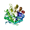
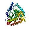
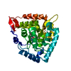
| Title | Crystal structure of endo-1,4-beta-mannanase from Eisenia fetida | |||||||||
 Components Components | endo-1,4-beta-mannanase | |||||||||
 Keywords Keywords | CARBOHYDRATE / cold adaptation / GH5 family | |||||||||
| Function / homology | Cellulase (glycosyl hydrolase family 5) / Glycosidases / Glycoside hydrolase superfamily / TIM Barrel / Alpha-Beta Barrel / Alpha Beta / ISOPROPYL ALCOHOL / Endo-1,4-beta-mannanase Function and homology information Function and homology information | |||||||||
| Biological species |  | |||||||||
| Method |  X-RAY DIFFRACTION / SYNCHROTRON / MOLECULAR REPLACEMENT / Resolution: 1.7 Å X-RAY DIFFRACTION / SYNCHROTRON / MOLECULAR REPLACEMENT / Resolution: 1.7 Å | |||||||||
 Authors Authors | Hirano, Y. / Ueda, M. / Tamada, T. | |||||||||
| Funding support |  Japan, 2items Japan, 2items
| |||||||||
 Citation Citation | Journal: Enzyme.Microb.Technol. / Year: 2018 Title: Gene cloning, expression, and X-ray crystallographic analysis of a beta-mannanase from Eisenia fetida. Authors: Ueda, M. / Hirano, Y. / Fukuhara, H. / Naka, Y. / Nakazawa, M. / Sakamoto, T. / Ogata, Y. / Tamada, T. | |||||||||
| History |
|
- Structure visualization
| Structure viewer | Molecule: MolmilJmol/JSmol |
|---|
- Downloads & links
-Download
| PDBx/mmCIF format | 5y6t.cif.gz | 92.9 KB | Display | PDBx/mmCIF format |
|---|---|---|---|---|
| PDB format | pdb5y6t.ent.gz | 67.5 KB | Display | PDB format |
| PDBx/mmJSON format | 5y6t.json.gz | Tree view | PDBx/mmJSON format | |
| Others |  Other downloads Other downloads |
-Validation report
| Arichive directory | https://data.pdbj.org/pub/pdb/validation_reports/y6/5y6tftp://data.pdbj.org/pub/pdb/validation_reports/y6/5y6t | HTTPS FTP |
|---|
-Related structure data
| Related structure data |  2c0hS S: Starting model for refinement |
|---|---|
| Similar structure data |


-Links
 PDBj
PDBj
- Assembly
| Deposited unit | 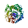
| ||||||||
|---|---|---|---|---|---|---|---|---|---|
| 1 |
| ||||||||
| Unit cell |
|
-Components
| #1: Protein | Mass: 42627.562 Da / Num. of mol.: 1 Source method: isolated from a genetically manipulated source Source: (gene. exp.)  Komagataella pastoris GS115 (fungus) / References: UniProt: A0A3B6UEQ6*PLUS Komagataella pastoris GS115 (fungus) / References: UniProt: A0A3B6UEQ6*PLUS |
|---|---|
| #2: Chemical | ChemComp-TRS /   Mass: 122.143 Da / Num. of mol.: 1 / Source method: obtained synthetically / Formula: C4H12NO3 / Comment: pH buffer*YM Mass: 122.143 Da / Num. of mol.: 1 / Source method: obtained synthetically / Formula: C4H12NO3 / Comment: pH buffer*YM |
| #3: Chemical | ChemComp-IPA /   Mass: 60.095 Da / Num. of mol.: 1 / Source method: obtained synthetically / Formula: C3H8O / Comment: alkaloid*YM Mass: 60.095 Da / Num. of mol.: 1 / Source method: obtained synthetically / Formula: C3H8O / Comment: alkaloid*YM |
| #4: Water | ChemComp-HOH /  Mass: 18.015 Da / Num. of mol.: 236 / Source method: isolated from a natural source / Formula: H2O Mass: 18.015 Da / Num. of mol.: 236 / Source method: isolated from a natural source / Formula: H2O |
| Has protein modification | Y |
-Experimental details
-Experiment
| Experiment | Method: X-RAY DIFFRACTION / Number of used crystals: 1 |
|---|
- Sample preparation
| Crystal | Density Matthews: 1.91 Å3/Da / Density % sol: 30.49 % |
|---|---|
| Crystal grow | Temperature: 293 K / Method: vapor diffusion, hanging drop / pH: 8.5 Details: 1 M ammonium citrate, 15%(v/v) iso-propanol, 0.1 M Tris-HCl pH 8.5 |
-Data collection
| Diffraction | Mean temperature: 100 K |
|---|---|
| Diffraction source | Source: SYNCHROTRON / Site: Photon Factory / Beamline: BL-17A / Wavelength: 0.98 Å |
| Detector | Type: DECTRIS PILATUS3 S 6M / Detector: PIXEL / Date: Dec 5, 2015 |
| Radiation | Monochromator: Si(111) / Protocol: SINGLE WAVELENGTH / Monochromatic (M) / Laue (L): M / Scattering type: x-ray |
| Radiation wavelength | Wavelength: 0.98 Å / Relative weight: 1 |
| Reflection | Resolution: 1.7→55 Å / Num. obs: 33115 / % possible obs: 98.4 % / Redundancy: 3.4 % / CC1/2: 0.998 / Rrim(I) all: 0.088 / Net I/σ(I): 13.1 |
| Reflection shell | Resolution: 1.7→1.81 Å / Redundancy: 3.3 % / Mean I/σ(I) obs: 2.7 / Num. unique obs: 10039 / CC1/2: 0.825 / Rrim(I) all: 0.554 / % possible all: 97.1 |
- Processing
| Software |
| ||||||||||||||||||||||||||||||||||||||||||||||||||||||||||||||||||||||||||||||||||||||||||||||||||||||||||||||||||||||||||||||||||||||||||||||||||||||||||||||||||||||||||||||||||||||
|---|---|---|---|---|---|---|---|---|---|---|---|---|---|---|---|---|---|---|---|---|---|---|---|---|---|---|---|---|---|---|---|---|---|---|---|---|---|---|---|---|---|---|---|---|---|---|---|---|---|---|---|---|---|---|---|---|---|---|---|---|---|---|---|---|---|---|---|---|---|---|---|---|---|---|---|---|---|---|---|---|---|---|---|---|---|---|---|---|---|---|---|---|---|---|---|---|---|---|---|---|---|---|---|---|---|---|---|---|---|---|---|---|---|---|---|---|---|---|---|---|---|---|---|---|---|---|---|---|---|---|---|---|---|---|---|---|---|---|---|---|---|---|---|---|---|---|---|---|---|---|---|---|---|---|---|---|---|---|---|---|---|---|---|---|---|---|---|---|---|---|---|---|---|---|---|---|---|---|---|---|---|---|---|
| Refinement | Method to determine structure: MOLECULAR REPLACEMENT Starting model: 2C0H Resolution: 1.7→54.2 Å / Cor.coef. Fo:Fc: 0.974 / Cor.coef. Fo:Fc free: 0.96 / SU B: 1.956 / SU ML: 0.065 / Cross valid method: THROUGHOUT / ESU R: 0.092 / ESU R Free: 0.093
| ||||||||||||||||||||||||||||||||||||||||||||||||||||||||||||||||||||||||||||||||||||||||||||||||||||||||||||||||||||||||||||||||||||||||||||||||||||||||||||||||||||||||||||||||||||||
| Solvent computation | Ion probe radii: 0.8 Å / Shrinkage radii: 0.8 Å / VDW probe radii: 1.2 Å | ||||||||||||||||||||||||||||||||||||||||||||||||||||||||||||||||||||||||||||||||||||||||||||||||||||||||||||||||||||||||||||||||||||||||||||||||||||||||||||||||||||||||||||||||||||||
| Displacement parameters | Biso mean: 23.891 Å2
| ||||||||||||||||||||||||||||||||||||||||||||||||||||||||||||||||||||||||||||||||||||||||||||||||||||||||||||||||||||||||||||||||||||||||||||||||||||||||||||||||||||||||||||||||||||||
| Refinement step | Cycle: 1 / Resolution: 1.7→54.2 Å
| ||||||||||||||||||||||||||||||||||||||||||||||||||||||||||||||||||||||||||||||||||||||||||||||||||||||||||||||||||||||||||||||||||||||||||||||||||||||||||||||||||||||||||||||||||||||
| Refine LS restraints |
|