 Movie
Movie Controller


+ Open data

- Basic information
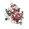
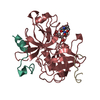
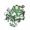
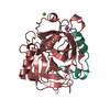
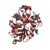
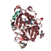
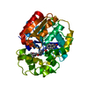
| Entry | Database: PDB / ID: 5y5d | ||||||
|---|---|---|---|---|---|---|---|
| Title | The crystal structure of VrEH2 mutant M263W | ||||||
 Components Components | Epoxide hydrolase | ||||||
 Keywords Keywords | HYDROLASE / Epoxide hydrolase | ||||||
| Function / homology |  Function and homology information Function and homology information | ||||||
| Biological species |  Vigna radiata (mung bean) Vigna radiata (mung bean) | ||||||
| Method |  X-RAY DIFFRACTION / SYNCHROTRON / MOLECULAR REPLACEMENT / Resolution: 1.85 Å X-RAY DIFFRACTION / SYNCHROTRON / MOLECULAR REPLACEMENT / Resolution: 1.85 Å | ||||||
 Authors Authors | Xu, J.H. / Yu, H.L. / Zhou, J.H. / Kong, X.D. / Li, F.L. | ||||||
 Citation Citation | Journal: To Be Published Title: The crystal structure of VrEH2 mutant M263W Authors: Xu, J.H. / Yu, H.L. / Zhou, J.H. / Kong, X.D. / Li, F.L. | ||||||
| History |
|
- Structure visualization
| Structure viewer | Molecule: MolmilJmol/JSmol |
|---|
- Downloads & links
-Download
| PDBx/mmCIF format | 5y5d.cif.gz | 85.6 KB | Display | PDBx/mmCIF format |
|---|---|---|---|---|
| PDB format | pdb5y5d.ent.gz | 61.7 KB | Display | PDB format |
| PDBx/mmJSON format | 5y5d.json.gz | Tree view | PDBx/mmJSON format | |
| Others |  Other downloads Other downloads |
-Validation report
| Arichive directory | https://data.pdbj.org/pub/pdb/validation_reports/y5/5y5dftp://data.pdbj.org/pub/pdb/validation_reports/y5/5y5d | HTTPS FTP |
|---|
-Related structure data
| Related structure data |  5xm6S S: Starting model for refinement |
|---|---|
| Similar structure data |
-Links
 PDBj
PDBj


- Assembly
| Deposited unit | 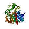
| ||||||||
|---|---|---|---|---|---|---|---|---|---|
| 1 |
| ||||||||
| Unit cell |
| ||||||||
| Components on special symmetry positions |
|
-Components
| #1: Protein | Mass: 37388.359 Da / Num. of mol.: 1 / Mutation: G3F, V4I, M263W Source method: isolated from a genetically manipulated source Source: (gene. exp.) Vigna radiata (mung bean) / Gene: EH2 / Production host:  |
|---|---|
| #2: Water | ChemComp-HOH /  Mass: 18.015 Da / Num. of mol.: 322 / Source method: isolated from a natural source / Formula: H2O Mass: 18.015 Da / Num. of mol.: 322 / Source method: isolated from a natural source / Formula: H2O |
-Experimental details
-Experiment
| Experiment | Method: X-RAY DIFFRACTION / Number of used crystals: 1 |
|---|
- Sample preparation
| Crystal | Density Matthews: 2.14 Å3/Da / Density % sol: 42.59 % / Description: rhombus |
|---|---|
| Crystal grow | Temperature: 285 K / Method: vapor diffusion, sitting drop / pH: 8.5 / Details: PEG3350, Tris-HCL, Ethylene glycol. |
-Data collection
| Diffraction | Mean temperature: 100 K |
|---|---|
| Diffraction source | Source: SYNCHROTRON / Site: SSRF  / Beamline: BL19U1 / Wavelength: 0.9791 Å / Beamline: BL19U1 / Wavelength: 0.9791 Å |
| Detector | Type: DECTRIS PILATUS3 6M / Detector: PIXEL / Date: Mar 10, 2017 |
| Radiation | Protocol: SINGLE WAVELENGTH / Monochromatic (M) / Laue (L): M / Scattering type: x-ray |
| Radiation wavelength | Wavelength: 0.9791 Å / Relative weight: 1 |
| Reflection | Resolution: 1.85→50 Å / Num. obs: 28594 / % possible obs: 99.7 % / Observed criterion σ(I): 0.9 / Redundancy: 24.5 % / Rmerge(I) obs: 0.091 / Rsym value: 0.091 / Net I/σ(I): 38.444 |
| Reflection shell | Resolution: 1.85→1.88 Å / Redundancy: 21.4 % / Rmerge(I) obs: 0.866 / Mean I/σ(I) obs: 2.5 / Num. unique obs: 28594 / Rsym value: 0.866 / % possible all: 96 |
- Processing
| Software |
| |||||||||||||||||||||||||||||||||||||||||||||||||||||||||||||||||||||||||||||
|---|---|---|---|---|---|---|---|---|---|---|---|---|---|---|---|---|---|---|---|---|---|---|---|---|---|---|---|---|---|---|---|---|---|---|---|---|---|---|---|---|---|---|---|---|---|---|---|---|---|---|---|---|---|---|---|---|---|---|---|---|---|---|---|---|---|---|---|---|---|---|---|---|---|---|---|---|---|---|
| Refinement | Method to determine structure: MOLECULAR REPLACEMENT Starting model: 5XM6 Resolution: 1.85→25.422 Å / SU ML: 0.19 / Cross valid method: FREE R-VALUE / σ(F): 1.35 / Phase error: 24.29
| |||||||||||||||||||||||||||||||||||||||||||||||||||||||||||||||||||||||||||||
| Solvent computation | Shrinkage radii: 0.9 Å / VDW probe radii: 1.11 Å | |||||||||||||||||||||||||||||||||||||||||||||||||||||||||||||||||||||||||||||
| Refinement step | Cycle: LAST / Resolution: 1.85→25.422 Å
| |||||||||||||||||||||||||||||||||||||||||||||||||||||||||||||||||||||||||||||
| Refine LS restraints |
| |||||||||||||||||||||||||||||||||||||||||||||||||||||||||||||||||||||||||||||
| LS refinement shell |
|
