 Movie
Movie Controller


+ Open data

- Basic information
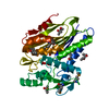
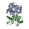
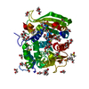
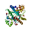
| Entry | Database: PDB / ID: 5xxa | ||||||
|---|---|---|---|---|---|---|---|
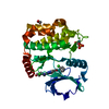
| Title | beta-1,4-mannanase-SeMet-RmMan134A | ||||||
 Components Components | endo-1,4-beta-mannanase | ||||||
 Keywords Keywords | HYDROLASE / protocolSeleno-methionine | ||||||
| Function / homology | : / Glycosyl hydrolase family 134 / Uncharacterized protein Function and homology information Function and homology information | ||||||
| Biological species |  Rhizopus microsporus (fungus) Rhizopus microsporus (fungus) | ||||||
| Method |  X-RAY DIFFRACTION / SYNCHROTRON / MOLECULAR REPLACEMENT / Resolution: 1.76 Å X-RAY DIFFRACTION / SYNCHROTRON / MOLECULAR REPLACEMENT / Resolution: 1.76 Å | ||||||
 Authors Authors | Jiang, Z.Q. / You, X. / Yang, D. / Huang, P. | ||||||
 Citation Citation | Journal: To Be Published Title: Structure of endo-1,4-beta-mannanase at 1.76 Angstroms resolution. Authors: Jiang, Z.Q. / You, X. / Yang, D. / Huang, P. | ||||||
| History |
|
- Structure visualization
| Structure viewer | Molecule: MolmilJmol/JSmol |
|---|
- Downloads & links
-Download
| PDBx/mmCIF format | 5xxa.cif.gz | 81.2 KB | Display | PDBx/mmCIF format |
|---|---|---|---|---|
| PDB format | pdb5xxa.ent.gz | 60.9 KB | Display | PDB format |
| PDBx/mmJSON format | 5xxa.json.gz | Tree view | PDBx/mmJSON format | |
| Others |  Other downloads Other downloads |
-Validation report
| Arichive directory | https://data.pdbj.org/pub/pdb/validation_reports/xx/5xxaftp://data.pdbj.org/pub/pdb/validation_reports/xx/5xxa | HTTPS FTP |
|---|
-Related structure data
| Similar structure data |
|---|


-Links
 PDBj
PDBj- Assembly
| Deposited unit | 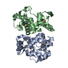
| ||||||||
|---|---|---|---|---|---|---|---|---|---|
| 1 |
| ||||||||
| Unit cell |
|
-Components
| #1: Protein | Mass: 18380.725 Da / Num. of mol.: 2 / Fragment: UNP residues 20-181 Source method: isolated from a genetically manipulated source Source: (gene. exp.) Rhizopus microsporus (fungus) / Gene: BCV71DRAFT_26579, RMCBS344292_04886 / Production host:  #2: Water | ChemComp-HOH / |  Mass: 18.015 Da / Num. of mol.: 417 / Source method: isolated from a natural source / Formula: H2O Mass: 18.015 Da / Num. of mol.: 417 / Source method: isolated from a natural source / Formula: H2OHas protein modification | Y | |
|---|
-Experimental details
-Experiment
| Experiment | Method: X-RAY DIFFRACTION / Number of used crystals: 1 |
|---|
- Sample preparation
| Crystal | Density Matthews: 2.49 Å3/Da / Density % sol: 50.59 % |
|---|---|
| Crystal grow | Temperature: 293 K / Method: vapor diffusion, sitting drop / Details: Tris-Hcl, tri-Sodium Citrate |
-Data collection
| Diffraction | Mean temperature: 100 K |
|---|---|
| Diffraction source | Source: SYNCHROTRON / Site: SSRF  / Beamline: BL18U1 / Wavelength: 0.979 Å / Beamline: BL18U1 / Wavelength: 0.979 Å |
| Detector | Type: DECTRIS PILATUS3 S 6M / Detector: PIXEL / Date: Jun 20, 2016 |
| Radiation | Protocol: SINGLE WAVELENGTH / Monochromatic (M) / Laue (L): M / Scattering type: x-ray |
| Radiation wavelength | Wavelength: 0.979 Å / Relative weight: 1 |
| Reflection | Resolution: 1.76→42.81 Å / Num. obs: 36268 / % possible obs: 98 % / Redundancy: 6.8 % / Rmerge(I) obs: 0.086 / Net I/σ(I): 8.1 |
| Reflection shell | Resolution: 1.76→1.82 Å / Redundancy: 6.8 % / Mean I/σ(I) obs: 27 / % possible all: 98.5 |
- Processing
| Software |
| ||||||||||||||||
|---|---|---|---|---|---|---|---|---|---|---|---|---|---|---|---|---|---|
| Refinement | Method to determine structure: MOLECULAR REPLACEMENT / Resolution: 1.76→42.81 Å / Cross valid method: FREE R-VALUE
| ||||||||||||||||
| Refinement step | Cycle: LAST / Resolution: 1.76→42.81 Å
|
