 Movie
Movie Controller

[English] 日本語
 Yorodumi
- PDB-5xq0: Structural basis of kindlin-mediated integrin recognition and act... -
+ Open data

- Basic information
| Entry | Database: PDB / ID: 5xq0 | ||||||
|---|---|---|---|---|---|---|---|
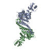
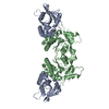
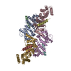
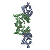
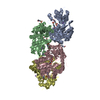
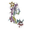
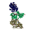
| Title | Structural basis of kindlin-mediated integrin recognition and activation | ||||||
 Components Components | Fermitin family homolog 2,Integrin beta-1 | ||||||
 Keywords Keywords | SIGNALING PROTEIN / Integrin Binding / Multi-domain containing protein | ||||||
| Function / homology |  Function and homology information Function and homology informationLocalization of the PINCH-ILK-PARVIN complex to focal adhesions / Laminin interactions / MET interacts with TNS proteins / protein transport within lipid bilayer / Elastic fibre formation / Cell-extracellular matrix interactions / Syndecan interactions / C-X3-C chemokine binding / Signal transduction by L1 / Molecules associated with elastic fibres ...Localization of the PINCH-ILK-PARVIN complex to focal adhesions / Laminin interactions / MET interacts with TNS proteins / protein transport within lipid bilayer / Elastic fibre formation / Cell-extracellular matrix interactions / Syndecan interactions / C-X3-C chemokine binding / Signal transduction by L1 / Molecules associated with elastic fibres / Fibronectin matrix formation / negative regulation of cell projection organization / MET activates PTK2 signaling / Basigin interactions / RAC3 GTPase cycle / ECM proteoglycans / TGF-beta receptor signaling activates SMADs / RAC2 GTPase cycle / integrin alpha3-beta1 complex / integrin alpha6-beta1 complex / integrin alpha7-beta1 complex / integrin alpha10-beta1 complex / integrin alpha11-beta1 complex / positive regulation of glutamate uptake involved in transmission of nerve impulse / adherens junction maintenance / integrin alpha5-beta1 complex / integrin alpha9-beta1 complex / regulation of collagen catabolic process / integrin alpha1-beta1 complex / integrin binding involved in cell-matrix adhesion / integrin alpha4-beta1 complex / hemidesmosome / collagen binding involved in cell-matrix adhesion / integrin alpha2-beta1 complex / RAC1 GTPase cycle / cell-cell adhesion mediated by integrin / formation of radial glial scaffolds / protein localization to cell junction / Turbulent (oscillatory, disturbed) flow shear stress activates signaling by PIEZO1 and integrins in endothelial cells / cerebellar climbing fiber to Purkinje cell synapse / Integrin cell surface interactions / Cell surface interactions at the vascular wall / positive regulation of mesenchymal stem cell proliferation / positive regulation of wound healing, spreading of epidermal cells / CD40 signaling pathway / calcium-independent cell-matrix adhesion / RHOG GTPase cycle / positive regulation of fibroblast growth factor receptor signaling pathway / GPER1 signaling / myelin sheath abaxonal region / reactive gliosis / integrin alphav-beta1 complex / basement membrane organization / regulation of synapse pruning / positive regulation of integrin activation / regulation of postsynaptic neurotransmitter receptor diffusion trapping / maintenance of postsynaptic specialization structure / bicellular tight junction assembly / tissue homeostasis / primordial germ cell migration / integrin activation / focal adhesion assembly / leukocyte tethering or rolling / type I transforming growth factor beta receptor binding / positive regulation of vascular endothelial growth factor signaling pathway / cell migration involved in sprouting angiogenesis / regulation of G protein-coupled receptor signaling pathway / myoblast fusion / positive regulation of cell-substrate adhesion / axon extension / cardiac muscle cell differentiation / myoblast differentiation / mesodermal cell differentiation / glycinergic synapse / regulation of cell morphogenesis / cell fate specification / Immunoregulatory interactions between a Lymphoid and a non-Lymphoid cell / cell projection organization / negative regulation of vascular permeability / positive regulation of fibroblast migration / negative regulation of Rho protein signal transduction / integrin complex / protein localization to membrane / cardiac muscle tissue development / cell adhesion mediated by integrin / regulation of spontaneous synaptic transmission / heterotypic cell-cell adhesion / I band / lamellipodium assembly / negative regulation of vasoconstriction / muscle organ development / sarcomere organization / leukocyte cell-cell adhesion / dendrite morphogenesis / limb development / negative regulation of neuron differentiation / positive regulation of wound healing / response to muscle activity / negative regulation of fat cell differentiation / cell-substrate adhesion Similarity search - Function | ||||||
| Biological species |  | ||||||
| Method |  X-RAY DIFFRACTION / SYNCHROTRON / MOLECULAR REPLACEMENT / Resolution: 2.75 Å X-RAY DIFFRACTION / SYNCHROTRON / MOLECULAR REPLACEMENT / Resolution: 2.75 Å | ||||||
 Authors Authors | Li, H. / Yang, H. / Sun, K. / Zhang, Z. / Yu, C. / Wei, Z. | ||||||
 Citation Citation | Journal: Proc. Natl. Acad. Sci. U.S.A. / Year: 2017 Title: Structural basis of kindlin-mediated integrin recognition and activation Authors: Li, H. / Deng, Y. / Sun, K. / Yang, H. / Liu, J. / Wang, M. / Zhang, Z. / Lin, J. / Wu, C. / Wei, Z. / Yu, C. | ||||||
| History |
|
- Structure visualization
| Structure viewer | Molecule: MolmilJmol/JSmol |
|---|
- Downloads & links
-Download
| PDBx/mmCIF format | 5xq0.cif.gz | 353.1 KB | Display | PDBx/mmCIF format |
|---|---|---|---|---|
| PDB format | pdb5xq0.ent.gz | 285.6 KB | Display | PDB format |
| PDBx/mmJSON format | 5xq0.json.gz | Tree view | PDBx/mmJSON format | |
| Others |  Other downloads Other downloads |
-Validation report
| Arichive directory | https://data.pdbj.org/pub/pdb/validation_reports/xq/5xq0ftp://data.pdbj.org/pub/pdb/validation_reports/xq/5xq0 | HTTPS FTP |
|---|
-Related structure data
| Related structure data | 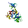 5xpyC  5xpzSC 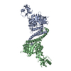 5xq1C S: Starting model for refinement C: citing same article ( |
|---|---|
| Similar structure data |

-Links
 PDBj
PDBj


- Assembly
| Deposited unit | 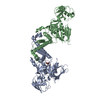
| ||||||||
|---|---|---|---|---|---|---|---|---|---|
| 1 |
| ||||||||
| Unit cell |
|
-Components
| #1: Protein | Mass: 56066.676 Da / Num. of mol.: 2 / Fragment: UNP residues 784-798 / Mutation: 168-217 deletion, 337-512 deletion Source method: isolated from a genetically manipulated source Details: Beta-integrin tail sequence was fused to the C-terminus of kindlin2 Source: (gene. exp.)  #2: Chemical | ChemComp-GOL / |   Mass: 92.094 Da / Num. of mol.: 1 / Source method: obtained synthetically / Formula: C3H8O3 Mass: 92.094 Da / Num. of mol.: 1 / Source method: obtained synthetically / Formula: C3H8O3#3: Water | ChemComp-HOH / |  Mass: 18.015 Da / Num. of mol.: 29 / Source method: isolated from a natural source / Formula: H2O Mass: 18.015 Da / Num. of mol.: 29 / Source method: isolated from a natural source / Formula: H2O |
|---|
-Experimental details
-Experiment
| Experiment | Method: X-RAY DIFFRACTION / Number of used crystals: 1 |
|---|
- Sample preparation
| Crystal | Density Matthews: 2.85 Å3/Da / Density % sol: 56.79 % |
|---|---|
| Crystal grow | Temperature: 289 K / Method: vapor diffusion, sitting drop / pH: 7.5 Details: 0.2 M potassium chloride, 0.05 M HEPES pH 7.5, 35% v/v pentaerythritol propoxylate |
-Data collection
| Diffraction | Mean temperature: 100 K | ||||||||||||||||||||||||||||||||||||||||||||||||||||||||||||||||||||||||||||||||||||||||||||||||||||||||||||||||||||||||||||||||||||||||||||||||||||||||||||||||||||||||
|---|---|---|---|---|---|---|---|---|---|---|---|---|---|---|---|---|---|---|---|---|---|---|---|---|---|---|---|---|---|---|---|---|---|---|---|---|---|---|---|---|---|---|---|---|---|---|---|---|---|---|---|---|---|---|---|---|---|---|---|---|---|---|---|---|---|---|---|---|---|---|---|---|---|---|---|---|---|---|---|---|---|---|---|---|---|---|---|---|---|---|---|---|---|---|---|---|---|---|---|---|---|---|---|---|---|---|---|---|---|---|---|---|---|---|---|---|---|---|---|---|---|---|---|---|---|---|---|---|---|---|---|---|---|---|---|---|---|---|---|---|---|---|---|---|---|---|---|---|---|---|---|---|---|---|---|---|---|---|---|---|---|---|---|---|---|---|---|---|---|
| Diffraction source | Source: SYNCHROTRON / Site: SSRF  / Beamline: BL17U1 / Wavelength: 0.98 Å / Beamline: BL17U1 / Wavelength: 0.98 Å | ||||||||||||||||||||||||||||||||||||||||||||||||||||||||||||||||||||||||||||||||||||||||||||||||||||||||||||||||||||||||||||||||||||||||||||||||||||||||||||||||||||||||
| Detector | Type: ADSC QUANTUM 315r / Detector: CCD / Date: Mar 22, 2015 | ||||||||||||||||||||||||||||||||||||||||||||||||||||||||||||||||||||||||||||||||||||||||||||||||||||||||||||||||||||||||||||||||||||||||||||||||||||||||||||||||||||||||
| Radiation | Protocol: SINGLE WAVELENGTH / Monochromatic (M) / Laue (L): M / Scattering type: x-ray | ||||||||||||||||||||||||||||||||||||||||||||||||||||||||||||||||||||||||||||||||||||||||||||||||||||||||||||||||||||||||||||||||||||||||||||||||||||||||||||||||||||||||
| Radiation wavelength | Wavelength: 0.98 Å / Relative weight: 1 | ||||||||||||||||||||||||||||||||||||||||||||||||||||||||||||||||||||||||||||||||||||||||||||||||||||||||||||||||||||||||||||||||||||||||||||||||||||||||||||||||||||||||
| Reflection | Resolution: 2.75→50 Å / Num. obs: 34443 / % possible obs: 98.7 % / Redundancy: 4 % / Biso Wilson estimate: 67.25 Å2 / Rmerge(I) obs: 0.086 / Rpim(I) all: 0.045 / Rrim(I) all: 0.098 / Χ2: 2.621 / Net I/σ(I): 12 / Num. measured all: 139053 | ||||||||||||||||||||||||||||||||||||||||||||||||||||||||||||||||||||||||||||||||||||||||||||||||||||||||||||||||||||||||||||||||||||||||||||||||||||||||||||||||||||||||
| Reflection shell | Diffraction-ID: 1
|
- Processing
| Software |
| ||||||||||||||||||||||||||||||||||||||||||||||||||||||||||||||||||||||||||||||||||||||||||||||||||||||||||||||||||||||||||||||||||||||||||||||||||||||||||||||||||||||||||||||||||||||||||||||||||||||||
|---|---|---|---|---|---|---|---|---|---|---|---|---|---|---|---|---|---|---|---|---|---|---|---|---|---|---|---|---|---|---|---|---|---|---|---|---|---|---|---|---|---|---|---|---|---|---|---|---|---|---|---|---|---|---|---|---|---|---|---|---|---|---|---|---|---|---|---|---|---|---|---|---|---|---|---|---|---|---|---|---|---|---|---|---|---|---|---|---|---|---|---|---|---|---|---|---|---|---|---|---|---|---|---|---|---|---|---|---|---|---|---|---|---|---|---|---|---|---|---|---|---|---|---|---|---|---|---|---|---|---|---|---|---|---|---|---|---|---|---|---|---|---|---|---|---|---|---|---|---|---|---|---|---|---|---|---|---|---|---|---|---|---|---|---|---|---|---|---|---|---|---|---|---|---|---|---|---|---|---|---|---|---|---|---|---|---|---|---|---|---|---|---|---|---|---|---|---|---|---|---|---|
| Refinement | Method to determine structure: MOLECULAR REPLACEMENT Starting model: 5XPZ Resolution: 2.75→45.858 Å / SU ML: 0.43 / Cross valid method: FREE R-VALUE / σ(F): 1.34 / Phase error: 27.8
| ||||||||||||||||||||||||||||||||||||||||||||||||||||||||||||||||||||||||||||||||||||||||||||||||||||||||||||||||||||||||||||||||||||||||||||||||||||||||||||||||||||||||||||||||||||||||||||||||||||||||
| Solvent computation | Shrinkage radii: 0.9 Å / VDW probe radii: 1.11 Å | ||||||||||||||||||||||||||||||||||||||||||||||||||||||||||||||||||||||||||||||||||||||||||||||||||||||||||||||||||||||||||||||||||||||||||||||||||||||||||||||||||||||||||||||||||||||||||||||||||||||||
| Displacement parameters | Biso max: 207.14 Å2 / Biso mean: 74.5935 Å2 / Biso min: 27.57 Å2 | ||||||||||||||||||||||||||||||||||||||||||||||||||||||||||||||||||||||||||||||||||||||||||||||||||||||||||||||||||||||||||||||||||||||||||||||||||||||||||||||||||||||||||||||||||||||||||||||||||||||||
| Refinement step | Cycle: final / Resolution: 2.75→45.858 Å
| ||||||||||||||||||||||||||||||||||||||||||||||||||||||||||||||||||||||||||||||||||||||||||||||||||||||||||||||||||||||||||||||||||||||||||||||||||||||||||||||||||||||||||||||||||||||||||||||||||||||||
| Refine LS restraints |
| ||||||||||||||||||||||||||||||||||||||||||||||||||||||||||||||||||||||||||||||||||||||||||||||||||||||||||||||||||||||||||||||||||||||||||||||||||||||||||||||||||||||||||||||||||||||||||||||||||||||||
| LS refinement shell | Refine-ID: X-RAY DIFFRACTION / Rfactor Rfree error: 0 / Total num. of bins used: 12
| ||||||||||||||||||||||||||||||||||||||||||||||||||||||||||||||||||||||||||||||||||||||||||||||||||||||||||||||||||||||||||||||||||||||||||||||||||||||||||||||||||||||||||||||||||||||||||||||||||||||||
| Refinement TLS params. | Method: refined / Refine-ID: X-RAY DIFFRACTION
| ||||||||||||||||||||||||||||||||||||||||||||||||||||||||||||||||||||||||||||||||||||||||||||||||||||||||||||||||||||||||||||||||||||||||||||||||||||||||||||||||||||||||||||||||||||||||||||||||||||||||
| Refinement TLS group |
|
