 Movie
Movie Controller

[English] 日本語
 Yorodumi
- PDB-5xo2: Crystal structure of human paired immunoglobulin-like type 2 rece... -
+ Open data

- Basic information
| Entry | Database: PDB / ID: 5xo2 | |||||||||
|---|---|---|---|---|---|---|---|---|---|---|
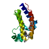
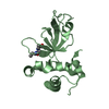
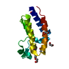
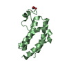
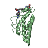
| Title | Crystal structure of human paired immunoglobulin-like type 2 receptor alpha with synthesized glycopeptide II | |||||||||
 Components Components |
| |||||||||
 Keywords Keywords | IMMUNE SYSTEM / membrane protein / immune receptor / viral entry inhibitor / glycopeptide | |||||||||
| Function / homology |  Function and homology information Function and homology informationhost cell Golgi membrane / MHC class I protein binding / host cell endosome membrane / Immunoregulatory interactions between a Lymphoid and a non-Lymphoid cell / viral envelope / symbiont entry into host cell / virion attachment to host cell / host cell plasma membrane / virion membrane / signal transduction ...host cell Golgi membrane / MHC class I protein binding / host cell endosome membrane / Immunoregulatory interactions between a Lymphoid and a non-Lymphoid cell / viral envelope / symbiont entry into host cell / virion attachment to host cell / host cell plasma membrane / virion membrane / signal transduction / extracellular exosome / identical protein binding / plasma membrane Similarity search - Function | |||||||||
| Biological species |  Homo sapiens (human) Homo sapiens (human)  Human herpesvirus 1 (Herpes simplex virus type 1) Human herpesvirus 1 (Herpes simplex virus type 1) | |||||||||
| Method |  X-RAY DIFFRACTION / SYNCHROTRON / MOLECULAR REPLACEMENT / Resolution: 2.201 Å X-RAY DIFFRACTION / SYNCHROTRON / MOLECULAR REPLACEMENT / Resolution: 2.201 Å | |||||||||
 Authors Authors | Furukawa, A. / Kakita, K. / Yamada, T. / Ishizuka, M. / Sakamoto, J. / Hatori, N. / Maeda, N. / Ohsaka, F. / Saitoh, T. / Nomura, T. ...Furukawa, A. / Kakita, K. / Yamada, T. / Ishizuka, M. / Sakamoto, J. / Hatori, N. / Maeda, N. / Ohsaka, F. / Saitoh, T. / Nomura, T. / Kuroki, K. / Nambu, H. / Arase, H. / Matsunaga, S. / Anada, M. / Ose, T. / Hashimoto, S. / Maenaka, K. | |||||||||
 Citation Citation | Journal: J. Biol. Chem. / Year: 2017 Title: Structural and thermodynamic analyses reveal critical features of glycopeptide recognition by the human PILR alpha immune cell receptor. Authors: Furukawa, A. / Kakita, K. / Yamada, T. / Ishizuka, M. / Sakamoto, J. / Hatori, N. / Maeda, N. / Ohsaka, F. / Saitoh, T. / Nomura, T. / Kuroki, K. / Nambu, H. / Arase, H. / Matsunaga, S. / ...Authors: Furukawa, A. / Kakita, K. / Yamada, T. / Ishizuka, M. / Sakamoto, J. / Hatori, N. / Maeda, N. / Ohsaka, F. / Saitoh, T. / Nomura, T. / Kuroki, K. / Nambu, H. / Arase, H. / Matsunaga, S. / Anada, M. / Ose, T. / Hashimoto, S. / Maenaka, K. | |||||||||
| History |
|
- Structure visualization
| Structure viewer | Molecule: MolmilJmol/JSmol |
|---|
- Downloads & links
-Download
| PDBx/mmCIF format | 5xo2.cif.gz | 68.2 KB | Display | PDBx/mmCIF format |
|---|---|---|---|---|
| PDB format | pdb5xo2.ent.gz | 48.5 KB | Display | PDB format |
| PDBx/mmJSON format | 5xo2.json.gz | Tree view | PDBx/mmJSON format | |
| Others |  Other downloads Other downloads |
-Validation report
| Arichive directory | https://data.pdbj.org/pub/pdb/validation_reports/xo/5xo2ftp://data.pdbj.org/pub/pdb/validation_reports/xo/5xo2 | HTTPS FTP |
|---|
-Related structure data
| Related structure data |  5xofC  3wv0S S: Starting model for refinement C: citing same article ( |
|---|---|
| Similar structure data |


-Links
 PDBj
PDBj- Assembly
| Deposited unit | 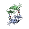
| ||||||||
|---|---|---|---|---|---|---|---|---|---|
| 1 | 
| ||||||||
| 2 | 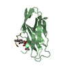
| ||||||||
| Unit cell |
|
-Components
| #1: Protein | Mass: 13967.697 Da / Num. of mol.: 2 / Fragment: UNP residues 32-150 Source method: isolated from a genetically manipulated source Source: (gene. exp.) Homo sapiens (human) / Gene: PILRA / Production host:  #2: Protein/peptide | Mass: 609.671 Da / Num. of mol.: 2 / Source method: obtained synthetically Source: (synth.) Human herpesvirus 1 (Herpes simplex virus type 1)References: UniProt: P06437 #3: Polysaccharide | Source method: isolated from a genetically manipulated source #4: Water | ChemComp-HOH / |  Mass: 18.015 Da / Num. of mol.: 49 / Source method: isolated from a natural source / Formula: H2O Mass: 18.015 Da / Num. of mol.: 49 / Source method: isolated from a natural source / Formula: H2OHas protein modification | Y | |
|---|
-Experimental details
-Experiment
| Experiment | Method: X-RAY DIFFRACTION / Number of used crystals: 1 |
|---|
- Sample preparation
| Crystal | Density Matthews: 2.28 Å3/Da / Density % sol: 46.12 % |
|---|---|
| Crystal grow | Temperature: 293 K / Method: vapor diffusion, sitting drop Details: 0.1M MES monohydrate pH 6.5, 1.6M Magnesium sulfate heptahydrate |
-Data collection
| Diffraction | Mean temperature: 100 K |
|---|---|
| Diffraction source | Source: SYNCHROTRON / Site: SPring-8  / Beamline: BL32XU / Wavelength: 1 Å / Beamline: BL32XU / Wavelength: 1 Å |
| Detector | Type: RAYONIX MX-225 / Detector: CCD / Date: Jul 13, 2015 |
| Radiation | Protocol: SINGLE WAVELENGTH / Monochromatic (M) / Laue (L): M / Scattering type: x-ray |
| Radiation wavelength | Wavelength: 1 Å / Relative weight: 1 |
| Reflection | Resolution: 2.2→50 Å / Num. obs: 13393 / % possible obs: 99.9 % / Redundancy: 3.8 % / Net I/σ(I): 25 |
| Reflection shell | Resolution: 2.2→2.24 Å / Redundancy: 3.6 % / Mean I/σ(I) obs: 8.4 / Num. unique all: 668 / % possible all: 99.7 |
- Processing
| Software |
| ||||||||||||||||
|---|---|---|---|---|---|---|---|---|---|---|---|---|---|---|---|---|---|
| Refinement | Method to determine structure: MOLECULAR REPLACEMENT Starting model: 3WV0 Resolution: 2.201→50 Å / Cross valid method: FREE R-VALUE
| ||||||||||||||||
| Refinement step | Cycle: LAST / Resolution: 2.201→50 Å
| ||||||||||||||||
| LS refinement shell | Resolution: 2.201→2.279 Å /
|
