 Movie
Movie Controller

[English] 日本語
 Yorodumi
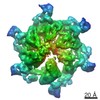
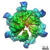
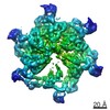
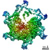
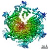
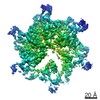
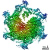
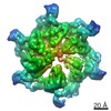
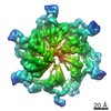
- PDB-5xmi: Cryo-EM Structure of the ATP-bound VPS4 mutant-E233Q hexamer (masked) -
+ Open data

- Basic information
| Entry | Database: PDB / ID: 5xmi | ||||||
|---|---|---|---|---|---|---|---|
| Title | Cryo-EM Structure of the ATP-bound VPS4 mutant-E233Q hexamer (masked) | ||||||
 Components Components | Vacuolar protein sorting-associated protein 4 | ||||||
 Keywords Keywords | PROTEIN TRANSPORT / ATPase / EESCRTIII / Vps4 | ||||||
| Function / homology |  Function and homology information Function and homology informationESCRT IV complex / Sealing of the nuclear envelope (NE) by ESCRT-III / late endosome to lysosome transport via multivesicular body sorting pathway / intralumenal vesicle formation / : / protein retention in Golgi apparatus / Endosomal Sorting Complex Required For Transport (ESCRT) / late endosome to vacuole transport via multivesicular body sorting pathway / sterol metabolic process / nuclear membrane reassembly ...ESCRT IV complex / Sealing of the nuclear envelope (NE) by ESCRT-III / late endosome to lysosome transport via multivesicular body sorting pathway / intralumenal vesicle formation / : / protein retention in Golgi apparatus / Endosomal Sorting Complex Required For Transport (ESCRT) / late endosome to vacuole transport via multivesicular body sorting pathway / sterol metabolic process / nuclear membrane reassembly / multivesicular body sorting pathway / vacuole organization / midbody abscission / membrane fission / multivesicular body assembly / late endosome to vacuole transport / ubiquitin-dependent protein catabolic process via the multivesicular body sorting pathway / plasma membrane repair / reticulophagy / endosomal transport / ATPase complex / nucleus organization / autophagosome maturation / nuclear pore / macroautophagy / autophagy / protein transport / midbody / endosome / endoplasmic reticulum / protein homodimerization activity / ATP hydrolysis activity / ATP binding / membrane / identical protein binding / plasma membrane / cytoplasm Similarity search - Function | ||||||
| Biological species |  | ||||||
| Method | ELECTRON MICROSCOPY / single particle reconstruction / cryo EM / Resolution: 3.9 Å | ||||||
 Authors Authors | Sun, S. / Li, L. / Yang, F. / Wang, X. / Fan, F. / Li, X. / Wang, H. / Sui, S. | ||||||
 Citation Citation | Journal: Nat Commun / Year: 2017 Title: Cryo-EM structures of the ATP-bound Vps4 hexamer and its complex with Vta1 at near-atomic resolution. Authors: Shan Sun / Lin Li / Fan Yang / Xiaojing Wang / Fenghui Fan / Mengyi Yang / Chunlai Chen / Xueming Li / Hong-Wei Wang / Sen-Fang Sui /  Abstract: The cellular ESCRT-III (endosomal sorting complex required for transport-III) and Vps4 (vacuolar protein sorting 4) comprise a common machinery that mediates a variety of membrane remodelling events. ...The cellular ESCRT-III (endosomal sorting complex required for transport-III) and Vps4 (vacuolar protein sorting 4) comprise a common machinery that mediates a variety of membrane remodelling events. Vps4 is essential for the machinery function by using the energy from ATP hydrolysis to disassemble the ESCRT-III polymer into individual proteins. Here, we report the structures of the ATP-bound Vps4 hexamer and its complex with the cofactor Vta1 (vps twenty associated 1) at resolutions of 3.9 and 4.2 Å, respectively, determined by electron cryo-microscopy. Six Vps4 subunits in both assemblies exhibit a spiral-shaped ring-like arrangement. Locating at the periphery of the hexameric ring, Vta1 dimer bridges two adjacent Vps4 subunits by two different interaction modes to promote the formation of the active Vps4 hexamer during ESCRT-III filament disassembly. The structural findings, together with the structure-guided biochemical and single-molecule analyses, provide important insights into the process of the ESCRT-III polymer disassembly by Vps4. | ||||||
| History |
|
- Structure visualization
| Movie |
Movie viewer |
|---|---|
| Structure viewer | Molecule: MolmilJmol/JSmol |
- Downloads & links
-Download
| PDBx/mmCIF format | 5xmi.cif.gz | 376.6 KB | Display | PDBx/mmCIF format |
|---|---|---|---|---|
| PDB format | pdb5xmi.ent.gz | 303.7 KB | Display | PDB format |
| PDBx/mmJSON format | 5xmi.json.gz | Tree view | PDBx/mmJSON format | |
| Others |  Other downloads Other downloads |
-Validation report
| Arichive directory | https://data.pdbj.org/pub/pdb/validation_reports/xm/5xmiftp://data.pdbj.org/pub/pdb/validation_reports/xm/5xmi | HTTPS FTP |
|---|
-Related structure data
| Related structure data | 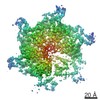 6733MC 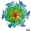 6734C 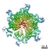 6735C 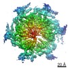 6736C 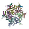 5xmkC M: map data used to model this data C: citing same article ( |
|---|---|
| Similar structure data |

-Links
 PDBj
PDBj


- Assembly
| Deposited unit | 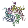
|
|---|---|
| 1 |
|
-Components
| #1: Protein | Mass: 48232.199 Da / Num. of mol.: 6 / Mutation: E233Q Source method: isolated from a genetically manipulated source Source: (gene. exp.) Strain: ATCC 204508 / S288c Gene: VPS4, CSC1, DID6, END13, GRD13, VPL4, VPT10, YPR173C, P9705.10 Production host:  #2: Chemical | ChemComp-ATP /   Mass: 507.181 Da / Num. of mol.: 5 / Source method: obtained synthetically / Formula: C10H16N5O13P3 / Comment: ATP, energy-carrying molecule*YM Mass: 507.181 Da / Num. of mol.: 5 / Source method: obtained synthetically / Formula: C10H16N5O13P3 / Comment: ATP, energy-carrying molecule*YM |
|---|
-Experimental details
-Experiment
| Experiment | Method: ELECTRON MICROSCOPY |
|---|---|
| EM experiment | Aggregation state: PARTICLE / 3D reconstruction method: single particle reconstruction |
- Sample preparation
| Component | Name: Vps4-E233Q hexamer / Type: ORGANELLE OR CELLULAR COMPONENT / Entity ID: #1 / Source: RECOMBINANT |
|---|---|
| Source (natural) | Organism: Strain: strain ATCC 204508 / S288c |
| Source (recombinant) | Organism: |
| Buffer solution | pH: 7.4 |
| Specimen | Embedding applied: NO / Shadowing applied: NO / Staining applied: NO / Vitrification applied: YES |
| Vitrification | Cryogen name: ETHANE |
- Electron microscopy imaging
| Experimental equipment |  Model: Titan Krios / Image courtesy: FEI Company |
|---|---|
| Microscopy | Model: FEI TITAN KRIOS |
| Electron gun | Electron source:  FIELD EMISSION GUN / Accelerating voltage: 300 kV / Illumination mode: OTHER FIELD EMISSION GUN / Accelerating voltage: 300 kV / Illumination mode: OTHER |
| Electron lens | Mode: BRIGHT FIELD |
| Image recording | Electron dose: 50 e/Å2 / Film or detector model: GATAN K2 SUMMIT (4k x 4k) |
- Processing
| Software | Name: REFMAC / Version: 5.8.0145 / Classification: refinement | ||||||||||||||||||||||||||||||||||||||||||||||||||||||||||||||||||||||||||||||||||||||||||||||||||||||||||
|---|---|---|---|---|---|---|---|---|---|---|---|---|---|---|---|---|---|---|---|---|---|---|---|---|---|---|---|---|---|---|---|---|---|---|---|---|---|---|---|---|---|---|---|---|---|---|---|---|---|---|---|---|---|---|---|---|---|---|---|---|---|---|---|---|---|---|---|---|---|---|---|---|---|---|---|---|---|---|---|---|---|---|---|---|---|---|---|---|---|---|---|---|---|---|---|---|---|---|---|---|---|---|---|---|---|---|---|
| CTF correction | Type: PHASE FLIPPING AND AMPLITUDE CORRECTION | ||||||||||||||||||||||||||||||||||||||||||||||||||||||||||||||||||||||||||||||||||||||||||||||||||||||||||
| 3D reconstruction | Resolution: 3.9 Å / Resolution method: FSC 0.143 CUT-OFF / Num. of particles: 106918 / Symmetry type: POINT | ||||||||||||||||||||||||||||||||||||||||||||||||||||||||||||||||||||||||||||||||||||||||||||||||||||||||||
| Refinement | Resolution: 3.9→166.32 Å / Cor.coef. Fo:Fc: 0.929 / SU B: 109.676 / SU ML: 1.366 Stereochemistry target values: MAXIMUM LIKELIHOOD WITH PHASES Details: HYDROGENS HAVE BEEN ADDED IN THE RIDING POSITIONS
| ||||||||||||||||||||||||||||||||||||||||||||||||||||||||||||||||||||||||||||||||||||||||||||||||||||||||||
| Solvent computation | Solvent model: PARAMETERS FOR MASK CACLULATION | ||||||||||||||||||||||||||||||||||||||||||||||||||||||||||||||||||||||||||||||||||||||||||||||||||||||||||
| Displacement parameters | Biso mean: 232.884 Å2
| ||||||||||||||||||||||||||||||||||||||||||||||||||||||||||||||||||||||||||||||||||||||||||||||||||||||||||
| Refinement step | Cycle: 1 / Total: 15000 | ||||||||||||||||||||||||||||||||||||||||||||||||||||||||||||||||||||||||||||||||||||||||||||||||||||||||||
| Refine LS restraints |
|