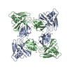
Deposited unit
A: Flagellar hook-associated protein 2
B: Flagellar hook-associated protein 2
C: Flagellar hook-associated protein 2
hetero molecules Summary Component details
Theoretical mass Number of molelcules Total (without water) 66,049 12 Polymers 65,460 3 Non-polymers 589 9 Water 0 0
1
A: Flagellar hook-associated protein 2
B: Flagellar hook-associated protein 2
hetero molecules
A: Flagellar hook-associated protein 2
B: Flagellar hook-associated protein 2
hetero molecules Summary Component details Symmetry operations Calculated values
Theoretical mass Number of molelcules Total (without water) 88,065 16 Polymers 87,280 4 Non-polymers 785 12 Water 0
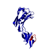
Type Name Symmetry operation Number identity operation 1_555 x,y,z 1 crystal symmetry operation 5_455 -x-1,y,-z 1
Buried area 7330 Å2 ΔGint -301 kcal/mol Surface area 33690 Å2 Method
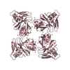
2
C: Flagellar hook-associated protein 2
hetero molecules
C: Flagellar hook-associated protein 2
hetero molecules
C: Flagellar hook-associated protein 2
hetero molecules
C: Flagellar hook-associated protein 2
hetero molecules Summary Component details Symmetry operations Calculated values
Theoretical mass Number of molelcules Total (without water) 88,065 16 Polymers 87,280 4 Non-polymers 785 12 Water 0
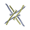
Type Name Symmetry operation Number identity operation 1_555 x,y,z 1 crystal symmetry operation 2_445 -x-1,-y-1,z 1 crystal symmetry operation 3_455 -y-1,x,z 1 crystal symmetry operation 4_545 y,-x-1,z 1
Buried area 7460 Å2 ΔGint -317 kcal/mol Surface area 32390 Å2 Method
Unit cell Length a, b, c (Å) 121.362, 121.362, 249.266 Angle α, β, γ (deg.) 90.00, 90.00, 90.00 Int Tables number 97 Space group name H-M I422
Components on special symmetry positions ID Model Components 1 1 C -301-ZN
Noncrystallographic symmetry (NCS) NCS domain ID Ens-ID Details (eV)1 1 A2 1 B3 1 C1 2 A2 2 B3 2 C
NCS domain segments Dom-ID Component-ID Ens-ID Refine code Auth asym-ID Auth seq-ID 1 1 1 3 A79 - 103 2 1 1 3 B79 - 103 3 1 1 3 C79 - 103 1 2 1 3 A226 - 299 2 2 1 3 B226 - 299 3 2 1 3 C226 - 299 1 1 2 3 A104 - 199 2 1 2 3 B104 - 199 3 1 2 3 C104 - 199 1 2 2 3 A203 - 225 2 2 2 3 B203 - 225 3 2 2 3 C203 - 225
NCS ensembles
 Movie
Movie Controller

 Yorodumi
 Open data
 Basic information
 Components
 Keywords
 Function and homology information
 Serratia marcescens (bacteria)
 X-RAY DIFFRACTION /
  Authors
 Citation
 Structure visualization
 Downloads & links
 Other downloads
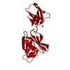


 PDBj
PDBj Assembly
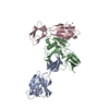

 Mass: 65.409 Da / Num. of mol.: 9 / Source method: obtained synthetically / Formula: Zn
 Sample preparation
 / Beamline: 7A (6B, 6C1) / Wavelength: 1.00004 Å
 Processing