specification of floral organ number / mucilage extrusion from seed coat / specification of floral organ identity / seed coat development / plant ovule development / flower development / cell fate specification / DNA-binding transcription factor activity, RNA polymerase II-specific / protein dimerization activity / RNA polymerase II cis-regulatory region sequence-specific DNA binding ...specification of floral organ number / mucilage extrusion from seed coat / specification of floral organ identity / seed coat development / plant ovule development / flower development / cell fate specification / DNA-binding transcription factor activity, RNA polymerase II-specific / protein dimerization activity / RNA polymerase II cis-regulatory region sequence-specific DNA binding / DNA-binding transcription factor activity / regulation of transcription by RNA polymerase II / DNA-templated transcription / positive regulation of transcription by RNA polymerase II / DNA binding / nucleus Similarity search - Function
Resolution: 2.49→44.5 Å / Cor.coef. Fo:Fc: 0.8791 / Cor.coef. Fo:Fc free: 0.8592 / Cross valid method: THROUGHOUT / σ(F): 0 Details: THE DATA WAS HIGHLY ANISOTROPIC WITH DIFFRACTION TO 2.49 ALONG A AND B AXIS AND 3.5 ALONG C AXIS. THIS ACCOUNTS FOR THE LOW COMPLETENESS IN THE HIGHEST RESOLUTION SHELLS. THE UCLA MBI SERVER ...Details: THE DATA WAS HIGHLY ANISOTROPIC WITH DIFFRACTION TO 2.49 ALONG A AND B AXIS AND 3.5 ALONG C AXIS. THIS ACCOUNTS FOR THE LOW COMPLETENESS IN THE HIGHEST RESOLUTION SHELLS. THE UCLA MBI SERVER WAS USED TO DETERMINE THE BEST RESOLUTION LIMITS ALONG THE DIFFERENT AXIS (M. STRONG, M.R. SAWAYA, S. WANG, M. PHILLIPS, D. CASCIO, D. EISENBERG, PROC NATL ACAD SCI USA. 103, 8060-8-65, 2006)
In the structure databanks used in Yorodumi, some data are registered as the other names, "COVID-19 virus" and "2019-nCoV". Here are the details of the virus and the list of structure data.
Jan 31, 2019. EMDB accession codes are about to change! (news from PDBe EMDB page)
EMDB accession codes are about to change! (news from PDBe EMDB page)
The allocation of 4 digits for EMDB accession codes will soon come to an end. Whilst these codes will remain in use, new EMDB accession codes will include an additional digit and will expand incrementally as the available range of codes is exhausted. The current 4-digit format prefixed with “EMD-” (i.e. EMD-XXXX) will advance to a 5-digit format (i.e. EMD-XXXXX), and so on. It is currently estimated that the 4-digit codes will be depleted around Spring 2019, at which point the 5-digit format will come into force.
The EM Navigator/Yorodumi systems omit the EMD- prefix.
Related info.:Q: What is EMD? / ID/Accession-code notation in Yorodumi/EM Navigator
Yorodumi is a browser for structure data from EMDB, PDB, SASBDB, etc.
This page is also the successor to EM Navigator detail page, and also detail information page/front-end page for Omokage search.
The word "yorodu" (or yorozu) is an old Japanese word meaning "ten thousand". "mi" (miru) is to see.
Related info.:EMDB / PDB / SASBDB / Comparison of 3 databanks / Yorodumi Search / Aug 31, 2016. New EM Navigator & Yorodumi / Yorodumi Papers / Jmol/JSmol / Function and homology information / Changes in new EM Navigator and Yorodumi
 Movie
Movie Controller

 Yorodumi
 Open data
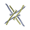
 Basic information
 Components
 Keywords
 Function and homology information
 X-RAY DIFFRACTION /
  Authors
 Citation
 Structure visualization
 Downloads & links
 Other downloads
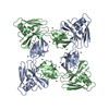
 PDBj
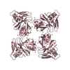
PDBj Assembly


 Mass: 18.015 Da / Num. of mol.: 262 / Source method: isolated from a natural source / Formula: H2O
 Sample preparation
 / Beamline: ID14-4 / Wavelength: 0.9393 Å
 Processing