 Movie
Movie Controller

[English] 日本語
 Yorodumi
- PDB-3u2r: Crystal structure of MarR transcription factor from Planctomyces ... -
+ Open data

- Basic information
| Entry | Database: PDB / ID: 3u2r | ||||||
|---|---|---|---|---|---|---|---|
| Title | Crystal structure of MarR transcription factor from Planctomyces limnophilus | ||||||
 Components Components | Regulatory protein MarR | ||||||
 Keywords Keywords | transcription regulator / Structural Genomics / PSI-Biology / Midwest Center for Structural Genomics / MCSG / helix-turn-helix | ||||||
| Function / homology |  Function and homology information Function and homology information | ||||||
| Biological species |  Planctomyces limnophilus (bacteria) Planctomyces limnophilus (bacteria) | ||||||
| Method |  X-RAY DIFFRACTION / SYNCHROTRON / SAD / Resolution: 2.2 Å X-RAY DIFFRACTION / SYNCHROTRON / SAD / Resolution: 2.2 Å | ||||||
 Authors Authors | Michalska, K. / Li, H. / Bearden, J. / Joachimiak, A. / Midwest Center for Structural Genomics (MCSG) | ||||||
 Citation Citation | Journal: To be Published Title: Crystal structure of MarR transcription factor from Planctomyces limnophilus Authors: Michalska, K. / Li, H. / Bearden, J. / Joachimiak, A. / Midwest Center for Structural Genomics (MCSG) | ||||||
| History |
|
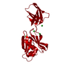
- Structure visualization
| Structure viewer | Molecule: MolmilJmol/JSmol |
|---|
- Downloads & links
-Download
| PDBx/mmCIF format | 3u2r.cif.gz | 70.5 KB | Display | PDBx/mmCIF format |
|---|---|---|---|---|
| PDB format | pdb3u2r.ent.gz | 52.4 KB | Display | PDB format |
| PDBx/mmJSON format | 3u2r.json.gz | Tree view | PDBx/mmJSON format | |
| Others |  Other downloads Other downloads |
-Validation report
| Arichive directory | https://data.pdbj.org/pub/pdb/validation_reports/u2/3u2rftp://data.pdbj.org/pub/pdb/validation_reports/u2/3u2r | HTTPS FTP |
|---|
-Related structure data
| Similar structure data | |
|---|---|
| Other databases |

-Links
 PDBj
PDBj- Assembly
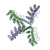
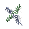
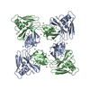
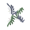
| Deposited unit | 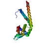
| ||||||||
|---|---|---|---|---|---|---|---|---|---|
| 1 | 
| ||||||||
| 2 | 
| ||||||||
| 3 | 
| ||||||||
| Unit cell |
|
-Components
| #1: Protein | Mass: 19897.035 Da / Num. of mol.: 1 Source method: isolated from a genetically manipulated source Source: (gene. exp.) Planctomyces limnophilus (bacteria) / Strain: DSM 3776 / Gene: Plim_0262 / Plasmid: PMCSG7 / Production host: |
|---|---|
| #2: Water | ChemComp-HOH /  Mass: 18.015 Da / Num. of mol.: 20 / Source method: isolated from a natural source / Formula: H2O Mass: 18.015 Da / Num. of mol.: 20 / Source method: isolated from a natural source / Formula: H2O |
| Has protein modification | Y |
-Experimental details
-Experiment
| Experiment | Method: X-RAY DIFFRACTION / Number of used crystals: 1 |
|---|
- Sample preparation
| Crystal | Density Matthews: 2.31 Å3/Da / Density % sol: 46.84 % |
|---|---|
| Crystal grow | Temperature: 289 K / Method: vapor diffusion, sitting drop / pH: 7 Details: 0.64 M malonic acid, 0.0875 M monoammonium malate, 0.14 M sodium acetate, 0.175 M sodium formate, 0.056 M ammonium tartrate, pH 7.0, 0.1 M Bis-Tris propane:NaOH, pH 7.0, VAPOR DIFFUSION, ...Details: 0.64 M malonic acid, 0.0875 M monoammonium malate, 0.14 M sodium acetate, 0.175 M sodium formate, 0.056 M ammonium tartrate, pH 7.0, 0.1 M Bis-Tris propane:NaOH, pH 7.0, VAPOR DIFFUSION, SITTING DROP, temperature 289K |
-Data collection
| Diffraction | Mean temperature: 100 K |
|---|---|
| Diffraction source | Source: SYNCHROTRON / Site: APS  / Beamline: 19-ID / Wavelength: 0.97929 Å / Beamline: 19-ID / Wavelength: 0.97929 Å |
| Detector | Type: ADSC QUANTUM 315r / Detector: CCD / Date: Jun 3, 2011 / Details: mirrors |
| Radiation | Monochromator: double crystal / Protocol: SINGLE WAVELENGTH / Monochromatic (M) / Laue (L): M / Scattering type: x-ray |
| Radiation wavelength | Wavelength: 0.97929 Å / Relative weight: 1 |
| Reflection | Resolution: 2.2→40 Å / Num. all: 10039 / Num. obs: 9939 / % possible obs: 99 % / Observed criterion σ(I): -3 / Redundancy: 5.9 % / Biso Wilson estimate: 48.26 Å2 / Rmerge(I) obs: 0.111 / Net I/σ(I): 13.4 |
| Reflection shell | Resolution: 2.2→2.24 Å / Redundancy: 5.8 % / Rmerge(I) obs: 0.619 / Mean I/σ(I) obs: 2.8 / Num. unique all: 993 / % possible all: 100 |
- Processing
| Software |
| |||||||||||||||||||||||||||||||||||||||||||||||||||||||||||||||||||||||||||||||||||||||||||||||||||||||||||||||||||||||||||||
|---|---|---|---|---|---|---|---|---|---|---|---|---|---|---|---|---|---|---|---|---|---|---|---|---|---|---|---|---|---|---|---|---|---|---|---|---|---|---|---|---|---|---|---|---|---|---|---|---|---|---|---|---|---|---|---|---|---|---|---|---|---|---|---|---|---|---|---|---|---|---|---|---|---|---|---|---|---|---|---|---|---|---|---|---|---|---|---|---|---|---|---|---|---|---|---|---|---|---|---|---|---|---|---|---|---|---|---|---|---|---|---|---|---|---|---|---|---|---|---|---|---|---|---|---|---|---|
| Refinement | Method to determine structure: SAD / Resolution: 2.2→32.48 Å / Cor.coef. Fo:Fc: 0.9335 / Cor.coef. Fo:Fc free: 0.8937 / SU R Cruickshank DPI: 0.511 / Isotropic thermal model: isotropic / Cross valid method: THROUGHOUT / σ(F): 0 / Stereochemistry target values: ML Details: HYDROGEN ATOMS HAVE BEEN ADDED IN THE RIDING POSITIONS
| |||||||||||||||||||||||||||||||||||||||||||||||||||||||||||||||||||||||||||||||||||||||||||||||||||||||||||||||||||||||||||||
| Displacement parameters | Biso mean: 60.3 Å2
| |||||||||||||||||||||||||||||||||||||||||||||||||||||||||||||||||||||||||||||||||||||||||||||||||||||||||||||||||||||||||||||
| Refine analyze | Luzzati coordinate error obs: 0.303 Å | |||||||||||||||||||||||||||||||||||||||||||||||||||||||||||||||||||||||||||||||||||||||||||||||||||||||||||||||||||||||||||||
| Refinement step | Cycle: LAST / Resolution: 2.2→32.48 Å
| |||||||||||||||||||||||||||||||||||||||||||||||||||||||||||||||||||||||||||||||||||||||||||||||||||||||||||||||||||||||||||||
| Refine LS restraints |
| |||||||||||||||||||||||||||||||||||||||||||||||||||||||||||||||||||||||||||||||||||||||||||||||||||||||||||||||||||||||||||||
| LS refinement shell | Resolution: 2.2→2.46 Å / Total num. of bins used: 5
| |||||||||||||||||||||||||||||||||||||||||||||||||||||||||||||||||||||||||||||||||||||||||||||||||||||||||||||||||||||||||||||
| Refinement TLS params. | Method: refined / Refine-ID: X-RAY DIFFRACTION
| |||||||||||||||||||||||||||||||||||||||||||||||||||||||||||||||||||||||||||||||||||||||||||||||||||||||||||||||||||||||||||||
| Refinement TLS group |
|
