 Movie
Movie Controller

[English] 日本語
 Yorodumi
- PDB-5xit: Crystal structure of RNF168 UDM1 in complex with Lys63-linked diu... -
+ Open data

- Basic information
| Entry | Database: PDB / ID: 5xit | ||||||
|---|---|---|---|---|---|---|---|
| Title | Crystal structure of RNF168 UDM1 in complex with Lys63-linked diubiquitin, form II | ||||||
 Components Components |
| ||||||
 Keywords Keywords | TRANSFERASE/RIBOSOMAL PROTEIN / protein complex / DNA repair / TRANSFERASE-RIBOSOMAL PROTEIN complex | ||||||
| Function / homology |  Function and homology information Function and homology informationFormation of the ternary complex, and subsequently, the 43S complex / APC/C:Cdc20 mediated degradation of Cyclin B / APC-Cdc20 mediated degradation of Nek2A / ER Quality Control Compartment (ERQC) / Regulation of PTEN localization / Downregulation of ERBB2:ERBB3 signaling / IRAK2 mediated activation of TAK1 complex / SMAD2/SMAD3:SMAD4 heterotrimer regulates transcription / PTK6 Regulates RTKs and Their Effectors AKT1 and DOK1 / Gap-filling DNA repair synthesis and ligation in GG-NER ...Formation of the ternary complex, and subsequently, the 43S complex / APC/C:Cdc20 mediated degradation of Cyclin B / APC-Cdc20 mediated degradation of Nek2A / ER Quality Control Compartment (ERQC) / Regulation of PTEN localization / Downregulation of ERBB2:ERBB3 signaling / IRAK2 mediated activation of TAK1 complex / SMAD2/SMAD3:SMAD4 heterotrimer regulates transcription / PTK6 Regulates RTKs and Their Effectors AKT1 and DOK1 / Gap-filling DNA repair synthesis and ligation in GG-NER / Fanconi Anemia Pathway / Endosomal Sorting Complex Required For Transport (ESCRT) / Negative regulation of FLT3 / Synthesis of active ubiquitin: roles of E1 and E2 enzymes / Regulation of expression of SLITs and ROBOs / IRAK1 recruits IKK complex / IRAK1 recruits IKK complex upon TLR7/8 or 9 stimulation / Downregulation of ERBB4 signaling / Stabilization of p53 / Formation of a pool of free 40S subunits / NOTCH3 Activation and Transmission of Signal to the Nucleus / Negative regulators of DDX58/IFIH1 signaling / Alpha-protein kinase 1 signaling pathway / Pexophagy / Regulation of pyruvate metabolism / Downregulation of TGF-beta receptor signaling / TGF-beta receptor signaling in EMT (epithelial to mesenchymal transition) / JNK (c-Jun kinases) phosphorylation and activation mediated by activated human TAK1 / Translesion synthesis by REV1 / Negative regulation of FGFR3 signaling / Negative regulation of FGFR4 signaling / Translesion synthesis by POLK / Regulation of NF-kappa B signaling / Negative regulation of FGFR1 signaling / Negative regulation of FGFR2 signaling / Regulation of TP53 Activity through Methylation / SCF-beta-TrCP mediated degradation of Emi1 / SRP-dependent cotranslational protein targeting to membrane / NRIF signals cell death from the nucleus / Translesion synthesis by POLI / Major pathway of rRNA processing in the nucleolus and cytosol / Recognition of DNA damage by PCNA-containing replication complex / p75NTR recruits signalling complexes / Interferon alpha/beta signaling / Nonsense Mediated Decay (NMD) independent of the Exon Junction Complex (EJC) / Negative regulation of MAPK pathway / Spry regulation of FGF signaling / Regulation of TP53 Degradation / Translesion Synthesis by POLH / Activated NOTCH1 Transmits Signal to the Nucleus / Formation of TC-NER Pre-Incision Complex / Negative regulation of MET activity / E3 ubiquitin ligases ubiquitinate target proteins / TRAF6-mediated induction of TAK1 complex within TLR4 complex / IRAK2 mediated activation of TAK1 complex upon TLR7/8 or 9 stimulation / Termination of translesion DNA synthesis / Nonsense Mediated Decay (NMD) enhanced by the Exon Junction Complex (EJC) / Autodegradation of Cdh1 by Cdh1:APC/C / APC/C:Cdc20 mediated degradation of Securin / Senescence-Associated Secretory Phenotype (SASP) / Josephin domain DUBs / DNA Damage Recognition in GG-NER / Dual Incision in GG-NER / Ubiquitin-dependent degradation of Cyclin D / Regulation of TBK1, IKKε (IKBKE)-mediated activation of IRF3, IRF7 / histone H2AK15 ubiquitin ligase activity / Downregulation of SMAD2/3:SMAD4 transcriptional activity / AUF1 (hnRNP D0) binds and destabilizes mRNA / Downregulation of ERBB2 signaling / Dual incision in TC-NER / Oncogene Induced Senescence / Translation initiation complex formation / Ribosomal scanning and start codon recognition / Degradation of CRY and PER proteins / Cdc20:Phospho-APC/C mediated degradation of Cyclin A / N-glycan trimming in the ER and Calnexin/Calreticulin cycle / Assembly of the pre-replicative complex / CDK-mediated phosphorylation and removal of Cdc6 / TNFR1-induced NF-kappa-B signaling pathway / HDR through Homologous Recombination (HRR) / Gap-filling DNA repair synthesis and ligation in TC-NER / Metalloprotease DUBs / Formation of Incision Complex in GG-NER / Activation of IRF3, IRF7 mediated by TBK1, IKKε (IKBKE) / Regulation of BACH1 activity / EGFR downregulation / Autodegradation of the E3 ubiquitin ligase COP1 / G2/M Checkpoints / Degradation of AXIN / TCF dependent signaling in response to WNT / Asymmetric localization of PCP proteins / APC/C:Cdh1 mediated degradation of Cdc20 and other APC/C:Cdh1 targeted proteins in late mitosis/early G1 / PINK1-PRKN Mediated Mitophagy / Regulation of RUNX3 expression and activity / Regulation of FZD by ubiquitination / Regulation of RAS by GAPs / Regulation of PTEN stability and activity / Inactivation of CSF3 (G-CSF) signaling / L13a-mediated translational silencing of Ceruloplasmin expression / SCF(Skp2)-mediated degradation of p27/p21 Similarity search - Function | ||||||
| Biological species |   Homo sapiens (human) Homo sapiens (human) | ||||||
| Method |  X-RAY DIFFRACTION / SYNCHROTRON / MOLECULAR REPLACEMENT / Resolution: 2.25 Å X-RAY DIFFRACTION / SYNCHROTRON / MOLECULAR REPLACEMENT / Resolution: 2.25 Å | ||||||
 Authors Authors | Takahashi, T.S. / Sato, Y. / Fukai, S. | ||||||
 Citation Citation | Journal: Nat Commun / Year: 2018 Title: Structural insights into two distinct binding modules for Lys63-linked polyubiquitin chains in RNF168. Authors: Takahashi, T.S. / Hirade, Y. / Toma, A. / Sato, Y. / Yamagata, A. / Goto-Ito, S. / Tomita, A. / Nakada, S. / Fukai, S. | ||||||
| History |
|
- Structure visualization
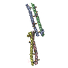
| Structure viewer | Molecule: MolmilJmol/JSmol |
|---|
- Downloads & links
-Download
| PDBx/mmCIF format | 5xit.cif.gz | 105.8 KB | Display | PDBx/mmCIF format |
|---|---|---|---|---|
| PDB format | pdb5xit.ent.gz | 80.4 KB | Display | PDB format |
| PDBx/mmJSON format | 5xit.json.gz | Tree view | PDBx/mmJSON format | |
| Others |  Other downloads Other downloads |
-Validation report
| Arichive directory | https://data.pdbj.org/pub/pdb/validation_reports/xi/5xitftp://data.pdbj.org/pub/pdb/validation_reports/xi/5xit | HTTPS FTP |
|---|
-Related structure data
| Related structure data |  5xisC 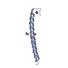 5xiuC  5ydkC  2fidS C: citing same article ( S: Starting model for refinement |
|---|---|
| Similar structure data |


-Links
 PDBj
PDBj


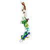
- Assembly
| Deposited unit | 
| ||||||||
|---|---|---|---|---|---|---|---|---|---|
| 1 | 
| ||||||||
| 2 | 
| ||||||||
| Unit cell |
|
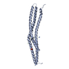
-Components
-Ubiquitin-40S ribosomal protein ... , 2 types, 4 molecules DFHB
| #1: Protein | Mass: 8604.845 Da / Num. of mol.: 2 / Mutation: K63R Source method: isolated from a genetically manipulated source Source: (gene. exp.)  #3: Protein | Mass: 8691.918 Da / Num. of mol.: 2 Source method: isolated from a genetically manipulated source Source: (gene. exp.) |
|---|
-Protein , 1 types, 2 molecules EA
| #2: Protein | Mass: 9618.506 Da / Num. of mol.: 2 / Fragment: UNP residues 113-188 Source method: isolated from a genetically manipulated source Source: (gene. exp.) Homo sapiens (human) / Gene: RNF168 / Plasmid: pCold-GST / Production host: References: UniProt: Q8IYW5, RING-type E3 ubiquitin transferase |
|---|
-Non-polymers , 3 types, 66 molecules 


| #4: Chemical | ChemComp-GOL /  Mass: 92.094 Da / Num. of mol.: 5 / Source method: obtained synthetically / Formula: C3H8O3 Mass: 92.094 Da / Num. of mol.: 5 / Source method: obtained synthetically / Formula: C3H8O3#5: Chemical |  Mass: 140.908 Da / Num. of mol.: 3 / Source method: obtained synthetically / Formula: Pr Mass: 140.908 Da / Num. of mol.: 3 / Source method: obtained synthetically / Formula: Pr#6: Water | ChemComp-HOH / | Mass: 18.015 Da / Num. of mol.: 58 / Source method: isolated from a natural source / Formula: H2O |
|---|
-Details
| Sequence details | additional C-terminal residue |
|---|
-Experimental details
-Experiment
| Experiment | Method: X-RAY DIFFRACTION / Number of used crystals: 1 |
|---|
- Sample preparation
| Crystal | Density Matthews: 2.39 Å3/Da / Density % sol: 48.58 % |
|---|---|
| Crystal grow | Temperature: 293 K / Method: vapor diffusion, hanging drop / pH: 6.5 Details: 23% PEG MME 2000, 0.1 M Bis-Tris (pH 6.5), 10 mM Pr acetate |
-Data collection
| Diffraction | Mean temperature: 100 K |
|---|---|
| Diffraction source | Source: SYNCHROTRON / Site: SPring-8  / Beamline: BL41XU / Wavelength: 1 Å / Beamline: BL41XU / Wavelength: 1 Å |
| Detector | Type: DECTRIS PILATUS3 6M / Detector: CMOS / Date: Oct 6, 2015 |
| Radiation | Protocol: SINGLE WAVELENGTH / Monochromatic (M) / Laue (L): M / Scattering type: x-ray |
| Radiation wavelength | Wavelength: 1 Å / Relative weight: 1 |
| Reflection | Resolution: 2.25→50 Å / Num. obs: 21397 / % possible obs: 90.2 % / Redundancy: 3.1 % / Rsym value: 0.112 / Net I/σ(I): 20.2 |
| Reflection shell | Resolution: 2.25→2.29 Å / Redundancy: 3 % / Mean I/σ(I) obs: 1.8 / Rsym value: 0.67 / % possible all: 91.5 |
- Processing
| Software |
| |||||||||||||||||||||||||||||||||||||||||||||||||||||||||||||||
|---|---|---|---|---|---|---|---|---|---|---|---|---|---|---|---|---|---|---|---|---|---|---|---|---|---|---|---|---|---|---|---|---|---|---|---|---|---|---|---|---|---|---|---|---|---|---|---|---|---|---|---|---|---|---|---|---|---|---|---|---|---|---|---|---|
| Refinement | Method to determine structure: MOLECULAR REPLACEMENT Starting model: 2FID Resolution: 2.25→46.986 Å / SU ML: 0.32 / Cross valid method: FREE R-VALUE / σ(F): 1.96 / Phase error: 31.26
| |||||||||||||||||||||||||||||||||||||||||||||||||||||||||||||||
| Solvent computation | Shrinkage radii: 0.9 Å / VDW probe radii: 1.11 Å | |||||||||||||||||||||||||||||||||||||||||||||||||||||||||||||||
| Refinement step | Cycle: LAST / Resolution: 2.25→46.986 Å
| |||||||||||||||||||||||||||||||||||||||||||||||||||||||||||||||
| Refine LS restraints |
| |||||||||||||||||||||||||||||||||||||||||||||||||||||||||||||||
| LS refinement shell |
|
