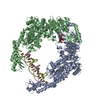
DNA BINDING PROTEIN / DNA Mismatch Repair / Sensor protein / AMPPnP
Function / homology
Function and homology information
mismatched DNA binding / ATP-dependent DNA damage sensor activity / mismatch repair / damaged DNA binding / ATP binding / cytosol Similarity search - Function
MutS, DNA mismatch repair protein; Chain A, domain 3 / MutS, DNA mismatch repair protein; Chain A, domain 3 - #10 / DNA repair protein MutS, domain I / DNA mismatch repair protein MutS / DNA mismatch repair protein MutS/MSH / DNA mismatch repair protein MutS-like, N-terminal / DNA mismatch repair protein MutS, connector domain / DNA mismatch repair protein MutS, clamp / DNA mismatch repair protein MutS, N-terminal / MutS, connector domain superfamily ...MutS, DNA mismatch repair protein; Chain A, domain 3 / MutS, DNA mismatch repair protein; Chain A, domain 3 - #10 / DNA repair protein MutS, domain I / DNA mismatch repair protein MutS / DNA mismatch repair protein MutS/MSH / DNA mismatch repair protein MutS-like, N-terminal / DNA mismatch repair protein MutS, connector domain / DNA mismatch repair protein MutS, clamp / DNA mismatch repair protein MutS, N-terminal / MutS, connector domain superfamily / MutS domain I / MutS domain II / MutS family domain IV / MutS domain III / DNA mismatch repair MutS family / DNA mismatch repair protein MutS, C-terminal / DNA mismatch repair protein MutS, core / DNA mismatch repair protein MutS, core domain superfamily / MutS domain V / DNA mismatch repair proteins mutS family signature. / DNA-binding domain of DNA mismatch repair MUTS family / ATPase domain of DNA mismatch repair MUTS family / MutS, DNA mismatch repair protein, domain I / P-loop containing nucleotide triphosphate hydrolases / Rossmann fold / P-loop containing nucleoside triphosphate hydrolase / Orthogonal Bundle / 3-Layer(aba) Sandwich / Mainly Alpha / Alpha Beta Similarity search - Domain/homology
Resolution: 3.3→49.93 Å / Cor.coef. Fo:Fc: 0.927 / Cor.coef. Fo:Fc free: 0.895 / SU B: 59.245 / SU ML: 0.45 / Cross valid method: THROUGHOUT / ESU R Free: 0.575 / Stereochemistry target values: MAXIMUM LIKELIHOOD / Details: HYDROGENS HAVE BEEN ADDED IN THE RIDING POSITIONS
Rfactor
Num. reflection
% reflection
Selection details
Rfree
0.27292
1646
5.1 %
RANDOM
Rwork
0.22126
-
-
-
obs
0.2239
30777
97.31 %
-
Solvent computation
Ion probe radii: 0.8 Å / Shrinkage radii: 0.8 Å / VDW probe radii: 1.2 Å / Solvent model: MASK
 Movie
Movie Controller


 Open data
 Basic information
 Components
 Keywords
 Function and homology information
 Neisseria gonorrhoeae (bacteria)
 X-RAY DIFFRACTION /
  Authors
 India, 1items
  Citation
 Structure visualization
 Downloads & links
 Other downloads
 PDBj
PDBj


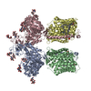
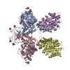
 Assembly

 Mass: 506.196 Da / Num. of mol.: 2 / Source method: obtained synthetically / Formula: C10H17N6O12P3 / Comment: AMP-PNP, energy-carrying molecule analogue*YM
 Sample preparation
 / Beamline: BM14 / Wavelength: 0.9537 Å
 Processing