 Movie
Movie Controller


+ Open data

- Basic information
| Entry | Database: PDB / ID: 5x9k | ||||||
|---|---|---|---|---|---|---|---|
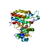
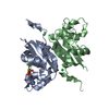
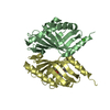
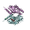
| Title | Structure of AusH from Aspergillus nidulans | ||||||
 Components Components | Austinol synthesis protein H | ||||||
 Keywords Keywords | ISOMERASE / meroterpenoid / Austinol | ||||||
| Function / homology | austinol biosynthetic process / dehydroaustinol biosynthetic process / : / terpenoid biosynthetic process / Austinoid biosynthesis clusters protein H Function and homology information Function and homology information | ||||||
| Biological species |  | ||||||
| Method |  X-RAY DIFFRACTION / SYNCHROTRON / MOLECULAR REPLACEMENT / Resolution: 1.798 Å X-RAY DIFFRACTION / SYNCHROTRON / MOLECULAR REPLACEMENT / Resolution: 1.798 Å | ||||||
 Authors Authors | Iwabuchi, T. / Matsuda, Y. / Abe, I. | ||||||
 Citation Citation | Journal: Nat. Chem. Biol. / Year: 2017 Title: Molecular basis for the unusual ring reconstruction in fungal meroterpenoid biogenesis Authors: Mori, T. / Iwabuchi, T. / Hoshino, S. / Wang, H. / Matsuda, Y. / Abe, I. | ||||||
| History |
|
- Structure visualization
| Structure viewer | Molecule: MolmilJmol/JSmol |
|---|
- Downloads & links
-Download
| PDBx/mmCIF format | 5x9k.cif.gz | 77.1 KB | Display | PDBx/mmCIF format |
|---|---|---|---|---|
| PDB format | pdb5x9k.ent.gz | 56.1 KB | Display | PDB format |
| PDBx/mmJSON format | 5x9k.json.gz | Tree view | PDBx/mmJSON format | |
| Others |  Other downloads Other downloads |
-Validation report
| Arichive directory | https://data.pdbj.org/pub/pdb/validation_reports/x9/5x9kftp://data.pdbj.org/pub/pdb/validation_reports/x9/5x9k | HTTPS FTP |
|---|
-Related structure data
| Related structure data | 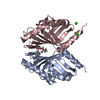 5wqfSC 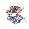 5wqgC 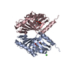 5wqhC 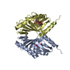 5wqiC 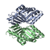 5x9jC S: Starting model for refinement C: citing same article ( |
|---|---|
| Similar structure data |


-Links
 PDBj
PDBj- Assembly
| Deposited unit | 
| ||||||||
|---|---|---|---|---|---|---|---|---|---|
| 1 |
| ||||||||
| Unit cell |
|
-Components
| #1: Protein | Mass: 18901.297 Da / Num. of mol.: 2 / Fragment: UNP residues 46-174 Source method: isolated from a genetically manipulated source Source: (gene. exp.)  #2: Water | ChemComp-HOH / |  Mass: 18.015 Da / Num. of mol.: 219 / Source method: isolated from a natural source / Formula: H2O Mass: 18.015 Da / Num. of mol.: 219 / Source method: isolated from a natural source / Formula: H2O |
|---|
-Experimental details
-Experiment
| Experiment | Method: X-RAY DIFFRACTION / Number of used crystals: 1 |
|---|
- Sample preparation
| Crystal | Density Matthews: 1.79 Å3/Da / Density % sol: 31.14 % |
|---|---|
| Crystal grow | Temperature: 293 K / Method: vapor diffusion, sitting drop Details: 100 mM Imidazole (pH 8.0), 10% PEG8000, 100 mM Ca(OAc)2 |
-Data collection
| Diffraction | Mean temperature: 100 K |
|---|---|
| Diffraction source | Source: SYNCHROTRON / Site: Photon Factory  / Beamline: AR-NE3A / Wavelength: 1 Å / Beamline: AR-NE3A / Wavelength: 1 Å |
| Detector | Type: DECTRIS PILATUS 2M-F / Detector: PIXEL / Date: Dec 16, 2015 |
| Radiation | Protocol: SINGLE WAVELENGTH / Monochromatic (M) / Laue (L): M / Scattering type: x-ray |
| Radiation wavelength | Wavelength: 1 Å / Relative weight: 1 |
| Reflection | Resolution: 1.798→50 Å / Num. obs: 25080 / % possible obs: 99.5 % / Redundancy: 3.3 % / Rmerge(I) obs: 0.058 / Net I/σ(I): 14.19 |
| Reflection shell | Resolution: 1.798→1.91 Å / Redundancy: 3.3 % / Rmerge(I) obs: 0.617 / Mean I/σ(I) obs: 1.85 / Num. unique obs: 3956 / % possible all: 98.4 |
- Processing
| Software |
| |||||||||||||||||||||||||||||||||||||||||||||||||||||||||||||||||||||||||||||||||||||||||||||||||||||||||
|---|---|---|---|---|---|---|---|---|---|---|---|---|---|---|---|---|---|---|---|---|---|---|---|---|---|---|---|---|---|---|---|---|---|---|---|---|---|---|---|---|---|---|---|---|---|---|---|---|---|---|---|---|---|---|---|---|---|---|---|---|---|---|---|---|---|---|---|---|---|---|---|---|---|---|---|---|---|---|---|---|---|---|---|---|---|---|---|---|---|---|---|---|---|---|---|---|---|---|---|---|---|---|---|---|---|---|
| Refinement | Method to determine structure: MOLECULAR REPLACEMENT Starting model: 5WQF Resolution: 1.798→47.055 Å / SU ML: 0.25 / Cross valid method: FREE R-VALUE / σ(F): 1.35 / Phase error: 26.61
| |||||||||||||||||||||||||||||||||||||||||||||||||||||||||||||||||||||||||||||||||||||||||||||||||||||||||
| Solvent computation | Shrinkage radii: 0.9 Å / VDW probe radii: 1.11 Å | |||||||||||||||||||||||||||||||||||||||||||||||||||||||||||||||||||||||||||||||||||||||||||||||||||||||||
| Refinement step | Cycle: LAST / Resolution: 1.798→47.055 Å
| |||||||||||||||||||||||||||||||||||||||||||||||||||||||||||||||||||||||||||||||||||||||||||||||||||||||||
| Refine LS restraints |
| |||||||||||||||||||||||||||||||||||||||||||||||||||||||||||||||||||||||||||||||||||||||||||||||||||||||||
| LS refinement shell |
|
