 Movie
Movie Controller

[English] 日本語
 Yorodumi
- PDB-5x6m: Crystal Structure of SMAD5-MH1 in complex with a composite DNA se... -
+ Open data

- Basic information
| Entry | Database: PDB / ID: 5x6m | |||||||||
|---|---|---|---|---|---|---|---|---|---|---|
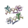
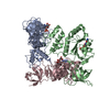
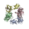
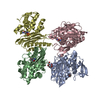
| Title | Crystal Structure of SMAD5-MH1 in complex with a composite DNA sequence | |||||||||
 Components Components |
| |||||||||
 Keywords Keywords | METAL BINDING PROTEIN/DNA / Smad5 MH1 domain / composite DNA / SBE DNA / GC-rich DNA / METAL BINDING PROTEIN-DNA complex | |||||||||
| Function / homology |  Function and homology information Function and homology informationnegative regulation of Fas signaling pathway / Mullerian duct regression / Signaling by BMP / osteoblast fate commitment / anti-Mullerian hormone receptor signaling pathway / heteromeric SMAD protein complex / DEAD/H-box RNA helicase binding / SMAD protein signal transduction / embryonic pattern specification / I-SMAD binding ...negative regulation of Fas signaling pathway / Mullerian duct regression / Signaling by BMP / osteoblast fate commitment / anti-Mullerian hormone receptor signaling pathway / heteromeric SMAD protein complex / DEAD/H-box RNA helicase binding / SMAD protein signal transduction / embryonic pattern specification / I-SMAD binding / cartilage development / ureteric bud development / germ cell development / anatomical structure morphogenesis / positive regulation of osteoblast differentiation / BMP signaling pathway / cardiac muscle contraction / transforming growth factor beta receptor signaling pathway / stem cell differentiation / erythrocyte differentiation / RNA polymerase II transcription regulatory region sequence-specific DNA binding / bone development / DNA-binding transcription repressor activity, RNA polymerase II-specific / sequence-specific double-stranded DNA binding / osteoblast differentiation / angiogenesis / intracellular iron ion homeostasis / DNA-binding transcription factor activity, RNA polymerase II-specific / cell differentiation / RNA polymerase II cis-regulatory region sequence-specific DNA binding / negative regulation of gene expression / ubiquitin protein ligase binding / regulation of transcription by RNA polymerase II / negative regulation of apoptotic process / negative regulation of transcription by RNA polymerase II / positive regulation of transcription by RNA polymerase II / protein-containing complex / mitochondrion / nucleoplasm / metal ion binding / nucleus / cytoplasm / cytosol Similarity search - Function | |||||||||
| Biological species |  synthetic construct (others) | |||||||||
| Method |  X-RAY DIFFRACTION / SYNCHROTRON / MOLECULAR REPLACEMENT / Resolution: 3.2 Å X-RAY DIFFRACTION / SYNCHROTRON / MOLECULAR REPLACEMENT / Resolution: 3.2 Å | |||||||||
 Authors Authors | Chai, N. / Wang, J. / Wang, Z.X. / Wu, J.W. | |||||||||
| Funding support |  China, 2items China, 2items
| |||||||||
 Citation Citation | Journal: Nucleic Acids Res. / Year: 2015 Title: Structural basis for the Smad5 MH1 domain to recognize different DNA sequences. Authors: Chai, N. / Li, W.X. / Wang, J. / Wang, Z.X. / Yang, S.M. / Wu, J.W. | |||||||||
| History |
|
- Structure visualization
| Structure viewer | Molecule: MolmilJmol/JSmol |
|---|
- Downloads & links
-Download
| PDBx/mmCIF format | 5x6m.cif.gz | 301.1 KB | Display | PDBx/mmCIF format |
|---|---|---|---|---|
| PDB format | pdb5x6m.ent.gz | 238.6 KB | Display | PDB format |
| PDBx/mmJSON format | 5x6m.json.gz | Tree view | PDBx/mmJSON format | |
| Others |  Other downloads Other downloads |
-Validation report
| Arichive directory | https://data.pdbj.org/pub/pdb/validation_reports/x6/5x6mftp://data.pdbj.org/pub/pdb/validation_reports/x6/5x6m | HTTPS FTP |
|---|
-Related structure data
| Related structure data |  5x6gC  5x6hC 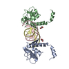 3kmpS C: citing same article ( S: Starting model for refinement |
|---|---|
| Similar structure data |


-Links
 PDBj
PDBj


- Assembly
| Deposited unit | 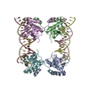
| ||||||||
|---|---|---|---|---|---|---|---|---|---|
| 1 |
| ||||||||
| Unit cell |
|
-Components
| #1: Protein | Mass: 17322.232 Da / Num. of mol.: 4 / Fragment: MH1 domain (UNP RESIDUES 1-143) Source method: isolated from a genetically manipulated source Source: (gene. exp.)  #2: DNA chain | Mass: 6736.367 Da / Num. of mol.: 2 / Source method: obtained synthetically / Source: (synth.) synthetic construct (others) #3: DNA chain | Mass: 6767.377 Da / Num. of mol.: 2 / Source method: obtained synthetically / Source: (synth.) synthetic construct (others) #4: Chemical | ChemComp-ZN /   Mass: 65.409 Da / Num. of mol.: 4 / Source method: obtained synthetically / Formula: Zn Mass: 65.409 Da / Num. of mol.: 4 / Source method: obtained synthetically / Formula: Zn |
|---|
-Experimental details
-Experiment
| Experiment | Method: X-RAY DIFFRACTION / Number of used crystals: 1 |
|---|
- Sample preparation
| Crystal | Density Matthews: 3.98 Å3/Da / Density % sol: 69.11 % |
|---|---|
| Crystal grow | Temperature: 277 K / Method: vapor diffusion, hanging drop / pH: 6.5 Details: 0.1M Bis-tris, pH 6.5, 0.2M LiNO3, 8% PEG 3350, 5% glycerol |
-Data collection
| Diffraction | Mean temperature: 100 K |
|---|---|
| Diffraction source | Source: SYNCHROTRON / Site: SSRF / Beamline: BL17U / Wavelength: 0.979065 Å |
| Detector | Type: ADSC QUANTUM 315r / Detector: CCD / Date: Nov 3, 2014 |
| Radiation | Monochromator: double crystal / Protocol: SINGLE WAVELENGTH / Monochromatic (M) / Laue (L): M / Scattering type: x-ray |
| Radiation wavelength | Wavelength: 0.979065 Å / Relative weight: 1 |
| Reflection | Resolution: 3.2→40 Å / Num. obs: 24244 / % possible obs: 98.8 % / Redundancy: 5.5 % / Rmerge(I) obs: 0.095 / Rpim(I) all: 0.046 / Net I/σ(I): 22.11 |
| Reflection shell | Resolution: 3.2→3.31 Å / Redundancy: 5.7 % / Rmerge(I) obs: 0.556 / Mean I/σ(I) obs: 4.55 / Num. unique obs: 2446 / CC1/2: 0.929 / Rpim(I) all: 0.257 / % possible all: 100 |
- Processing
| Software |
| |||||||||||||||||||||||||||||||||||||||||||||||||||||||||||||||||||||||||||||||||||||||||||||||||||||||||||||||||||||||||||||||||||||||||||||||||||||||||||||||||||||||||||||||||||||||||||||||||||||||||||||||||||||||||||||||||
|---|---|---|---|---|---|---|---|---|---|---|---|---|---|---|---|---|---|---|---|---|---|---|---|---|---|---|---|---|---|---|---|---|---|---|---|---|---|---|---|---|---|---|---|---|---|---|---|---|---|---|---|---|---|---|---|---|---|---|---|---|---|---|---|---|---|---|---|---|---|---|---|---|---|---|---|---|---|---|---|---|---|---|---|---|---|---|---|---|---|---|---|---|---|---|---|---|---|---|---|---|---|---|---|---|---|---|---|---|---|---|---|---|---|---|---|---|---|---|---|---|---|---|---|---|---|---|---|---|---|---|---|---|---|---|---|---|---|---|---|---|---|---|---|---|---|---|---|---|---|---|---|---|---|---|---|---|---|---|---|---|---|---|---|---|---|---|---|---|---|---|---|---|---|---|---|---|---|---|---|---|---|---|---|---|---|---|---|---|---|---|---|---|---|---|---|---|---|---|---|---|---|---|---|---|---|---|---|---|---|---|---|---|---|---|---|---|---|---|---|---|---|---|---|---|---|---|
| Refinement | Method to determine structure: MOLECULAR REPLACEMENT Starting model: 3KMP Resolution: 3.2→29.937 Å / SU ML: 0.36 / Cross valid method: FREE R-VALUE / σ(F): 1.96 / Phase error: 27.47
| |||||||||||||||||||||||||||||||||||||||||||||||||||||||||||||||||||||||||||||||||||||||||||||||||||||||||||||||||||||||||||||||||||||||||||||||||||||||||||||||||||||||||||||||||||||||||||||||||||||||||||||||||||||||||||||||||
| Solvent computation | Shrinkage radii: 0.9 Å / VDW probe radii: 1.11 Å | |||||||||||||||||||||||||||||||||||||||||||||||||||||||||||||||||||||||||||||||||||||||||||||||||||||||||||||||||||||||||||||||||||||||||||||||||||||||||||||||||||||||||||||||||||||||||||||||||||||||||||||||||||||||||||||||||
| Displacement parameters | Biso mean: 125.1 Å2 | |||||||||||||||||||||||||||||||||||||||||||||||||||||||||||||||||||||||||||||||||||||||||||||||||||||||||||||||||||||||||||||||||||||||||||||||||||||||||||||||||||||||||||||||||||||||||||||||||||||||||||||||||||||||||||||||||
| Refinement step | Cycle: LAST / Resolution: 3.2→29.937 Å
| |||||||||||||||||||||||||||||||||||||||||||||||||||||||||||||||||||||||||||||||||||||||||||||||||||||||||||||||||||||||||||||||||||||||||||||||||||||||||||||||||||||||||||||||||||||||||||||||||||||||||||||||||||||||||||||||||
| Refine LS restraints |
| |||||||||||||||||||||||||||||||||||||||||||||||||||||||||||||||||||||||||||||||||||||||||||||||||||||||||||||||||||||||||||||||||||||||||||||||||||||||||||||||||||||||||||||||||||||||||||||||||||||||||||||||||||||||||||||||||
| LS refinement shell |
| |||||||||||||||||||||||||||||||||||||||||||||||||||||||||||||||||||||||||||||||||||||||||||||||||||||||||||||||||||||||||||||||||||||||||||||||||||||||||||||||||||||||||||||||||||||||||||||||||||||||||||||||||||||||||||||||||
| Refinement TLS params. | Method: refined / Refine-ID: X-RAY DIFFRACTION
| |||||||||||||||||||||||||||||||||||||||||||||||||||||||||||||||||||||||||||||||||||||||||||||||||||||||||||||||||||||||||||||||||||||||||||||||||||||||||||||||||||||||||||||||||||||||||||||||||||||||||||||||||||||||||||||||||
| Refinement TLS group |
|
