 Movie
Movie Controller


+ Open data

- Basic information
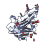
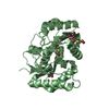
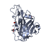
| Entry | Database: PDB / ID: 5x6a | ||||||
|---|---|---|---|---|---|---|---|
| Title | Crystal structure of an endoglucanase PMO-5 | ||||||
 Components Components | Endoglucanase, putative | ||||||
 Keywords Keywords | HYDROLASE / endoglucanase | ||||||
| Function / homology |  Function and homology information Function and homology informationlytic cellulose monooxygenase (C4-dehydrogenating) / cellulose catabolic process / monooxygenase activity / extracellular region Similarity search - Function | ||||||
| Biological species |  | ||||||
| Method |  X-RAY DIFFRACTION / SYNCHROTRON / MOLECULAR REPLACEMENT / Resolution: 1.7 Å X-RAY DIFFRACTION / SYNCHROTRON / MOLECULAR REPLACEMENT / Resolution: 1.7 Å | ||||||
 Authors Authors | Shen, Q. | ||||||
 Citation Citation | Journal: To Be Published Title: Crystal structure of an endoglucanase PMO-5 Authors: Shen, Q. | ||||||
| History |
|
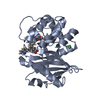
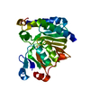
- Structure visualization
| Structure viewer | Molecule: MolmilJmol/JSmol |
|---|
- Downloads & links
-Download
| PDBx/mmCIF format | 5x6a.cif.gz | 109.3 KB | Display | PDBx/mmCIF format |
|---|---|---|---|---|
| PDB format | pdb5x6a.ent.gz | 82.7 KB | Display | PDB format |
| PDBx/mmJSON format | 5x6a.json.gz | Tree view | PDBx/mmJSON format | |
| Others |  Other downloads Other downloads |
-Validation report
| Summary document | 5x6a_validation.pdf.gz | 432.3 KB | Display | wwPDB validaton report |
|---|---|---|---|---|
| Full document | 5x6a_full_validation.pdf.gz | 434.7 KB | Display | |
| Data in XML | 5x6a_validation.xml.gz | 23.5 KB | Display | |
| Data in CIF | 5x6a_validation.cif.gz | 36.3 KB | Display | |
| Arichive directory | https://data.pdbj.org/pub/pdb/validation_reports/x6/5x6aftp://data.pdbj.org/pub/pdb/validation_reports/x6/5x6a | HTTPS FTP |
-Related structure data
| Related structure data | 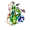 3zudS S: Starting model for refinement |
|---|---|
| Similar structure data |


-Links
 PDBj
PDBj- Assembly
| Deposited unit | 
| ||||||||
|---|---|---|---|---|---|---|---|---|---|
| 1 | 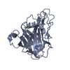
| ||||||||
| 2 | 
| ||||||||
| Unit cell |
|
-Components
| #1: Protein | Mass: 24171.695 Da / Num. of mol.: 2 Source method: isolated from a genetically manipulated source Source: (gene. exp.)  References: UniProt: Q4WP32, Hydrolases; Glycosylases; Glycosidases, i.e. enzymes that hydrolyse O- and S-glycosyl compounds #2: Chemical |   Mass: 24.305 Da / Num. of mol.: 2 / Source method: obtained synthetically / Formula: Mg Mass: 24.305 Da / Num. of mol.: 2 / Source method: obtained synthetically / Formula: Mg#3: Water | ChemComp-HOH / |  Mass: 18.015 Da / Num. of mol.: 534 / Source method: isolated from a natural source / Formula: H2O Mass: 18.015 Da / Num. of mol.: 534 / Source method: isolated from a natural source / Formula: H2OHas protein modification | Y | |
|---|
-Experimental details
-Experiment
| Experiment | Method: X-RAY DIFFRACTION / Number of used crystals: 1 |
|---|
- Sample preparation
| Crystal | Density Matthews: 2.27 Å3/Da / Density % sol: 45.91 % |
|---|---|
| Crystal grow | Temperature: 293 K / Method: vapor diffusion, hanging drop Details: 0.2M Magnesium chloride hexahydrate, 25% w/v Polyethylene glycol 3350, 0.1M BIS-TRIS pH 6.0 |
-Data collection
| Diffraction | Mean temperature: 77 K |
|---|---|
| Diffraction source | Source: SYNCHROTRON / Site: SSRF  / Beamline: BL17U / Wavelength: 0.9791 Å / Beamline: BL17U / Wavelength: 0.9791 Å |
| Detector | Type: DECTRIS PILATUS3 S 6M / Detector: PIXEL / Date: Jan 10, 2017 |
| Radiation | Protocol: SINGLE WAVELENGTH / Monochromatic (M) / Laue (L): M / Scattering type: x-ray |
| Radiation wavelength | Wavelength: 0.9791 Å / Relative weight: 1 |
| Reflection | Resolution: 1.7→27.5 Å / Num. obs: 46666 / % possible obs: 95.9 % / Redundancy: 3.6 % / Rmerge(I) obs: 0.117 / Rpim(I) all: 0.062 / Rsym value: 0.099 / Net I/σ(I): 37.3 |
| Reflection shell | Resolution: 1.7→1.73 Å / Redundancy: 3.8 % / Rmerge(I) obs: 0.476 / Mean I/σ(I) obs: 16.5 / Num. unique obs: 2194 / Rpim(I) all: 0.242 / Rsym value: 0.409 / % possible all: 95.5 |
- Processing
| Software |
| ||||||||||||||||||||||||||||||||||||||||||||||||||||||||||||||||||||||||||||||||||||||||||||||||||||||||||||||||||||||||||||||||||||||||||||||||||||||||||||||||||||||||||||||||||||||
|---|---|---|---|---|---|---|---|---|---|---|---|---|---|---|---|---|---|---|---|---|---|---|---|---|---|---|---|---|---|---|---|---|---|---|---|---|---|---|---|---|---|---|---|---|---|---|---|---|---|---|---|---|---|---|---|---|---|---|---|---|---|---|---|---|---|---|---|---|---|---|---|---|---|---|---|---|---|---|---|---|---|---|---|---|---|---|---|---|---|---|---|---|---|---|---|---|---|---|---|---|---|---|---|---|---|---|---|---|---|---|---|---|---|---|---|---|---|---|---|---|---|---|---|---|---|---|---|---|---|---|---|---|---|---|---|---|---|---|---|---|---|---|---|---|---|---|---|---|---|---|---|---|---|---|---|---|---|---|---|---|---|---|---|---|---|---|---|---|---|---|---|---|---|---|---|---|---|---|---|---|---|---|---|
| Refinement | Method to determine structure: MOLECULAR REPLACEMENT Starting model: 3zud Resolution: 1.7→27.5 Å / Cor.coef. Fo:Fc: 0.966 / Cor.coef. Fo:Fc free: 0.949 / SU B: 1.5 / SU ML: 0.051 / Cross valid method: THROUGHOUT / ESU R: 0.091 / ESU R Free: 0.088 / Details: HYDROGENS HAVE BEEN ADDED IN THE RIDING POSITIONS
| ||||||||||||||||||||||||||||||||||||||||||||||||||||||||||||||||||||||||||||||||||||||||||||||||||||||||||||||||||||||||||||||||||||||||||||||||||||||||||||||||||||||||||||||||||||||
| Solvent computation | Ion probe radii: 0.8 Å / Shrinkage radii: 0.8 Å / VDW probe radii: 1.2 Å | ||||||||||||||||||||||||||||||||||||||||||||||||||||||||||||||||||||||||||||||||||||||||||||||||||||||||||||||||||||||||||||||||||||||||||||||||||||||||||||||||||||||||||||||||||||||
| Displacement parameters | Biso mean: 12.114 Å2
| ||||||||||||||||||||||||||||||||||||||||||||||||||||||||||||||||||||||||||||||||||||||||||||||||||||||||||||||||||||||||||||||||||||||||||||||||||||||||||||||||||||||||||||||||||||||
| Refinement step | Cycle: 1 / Resolution: 1.7→27.5 Å
| ||||||||||||||||||||||||||||||||||||||||||||||||||||||||||||||||||||||||||||||||||||||||||||||||||||||||||||||||||||||||||||||||||||||||||||||||||||||||||||||||||||||||||||||||||||||
| Refine LS restraints |
|