 Movie
Movie Controller


+ Open data

- Basic information
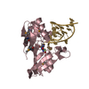
| Entry | Database: PDB / ID: 5x5v | ||||||
|---|---|---|---|---|---|---|---|
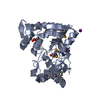
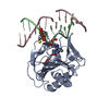
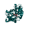
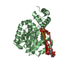
| Title | Crystal structure of pseudorabies virus glycoprotein D | ||||||
 Components Components | GD | ||||||
 Keywords Keywords | VIRAL PROTEIN / herpes simplex virus / pseudorabies virus / glycoprotein D | ||||||
| Function / homology | Herpesvirus glycoprotein D/GG/GX domain / Herpesvirus glycoprotein D/GG/GX domain / Immunoglobulin-like domain superfamily / membrane / GD Function and homology information Function and homology information | ||||||
| Biological species |  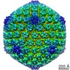 Suid herpesvirus 1 Suid herpesvirus 1 | ||||||
| Method |  X-RAY DIFFRACTION / SYNCHROTRON / MOLECULAR REPLACEMENT / Resolution: 1.5 Å X-RAY DIFFRACTION / SYNCHROTRON / MOLECULAR REPLACEMENT / Resolution: 1.5 Å | ||||||
 Authors Authors | Li, A. / Lu, G. / Qi, J. / Wu, L. / Tian, K. / Luo, T. / Shi, Y. / Yan, J. / Gao, G.F. | ||||||
 Citation Citation | Journal: To Be Published Title: Crystal structure of pseudorabies virus glycoprotein D Authors: Li, A. / Lu, G. / Qi, J. / Wu, L. / Tian, K. / Luo, T. / Shi, Y. / Yan, J. / Gao, G.F. | ||||||
| History |
|
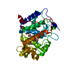
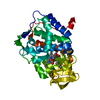
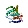
- Structure visualization
| Structure viewer | Molecule: MolmilJmol/JSmol |
|---|
- Downloads & links
-Download
| PDBx/mmCIF format | 5x5v.cif.gz | 127.8 KB | Display | PDBx/mmCIF format |
|---|---|---|---|---|
| PDB format | pdb5x5v.ent.gz | 97.6 KB | Display | PDB format |
| PDBx/mmJSON format | 5x5v.json.gz | Tree view | PDBx/mmJSON format | |
| Others |  Other downloads Other downloads |
-Validation report
| Arichive directory | https://data.pdbj.org/pub/pdb/validation_reports/x5/5x5vftp://data.pdbj.org/pub/pdb/validation_reports/x5/5x5v | HTTPS FTP |
|---|
-Related structure data
| Related structure data |  5x5wC 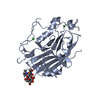 2c36S C: citing same article ( S: Starting model for refinement |
|---|---|
| Similar structure data |


-Links
 PDBj
PDBj- Assembly
| Deposited unit | 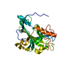
| |||||||||
|---|---|---|---|---|---|---|---|---|---|---|
| 1 |
| |||||||||
| 2 | 
| |||||||||
| Unit cell |
| |||||||||
| Components on special symmetry positions |
|
-Components
| #1: Protein | Mass: 44584.871 Da / Num. of mol.: 1 Source method: isolated from a genetically manipulated source Source: (gene. exp.) Suid herpesvirus 1 / Strain: Becker / Gene: US6 / Production host:  |
|---|---|
| #2: Water | ChemComp-HOH /  Mass: 18.015 Da / Num. of mol.: 334 / Source method: isolated from a natural source / Formula: H2O Mass: 18.015 Da / Num. of mol.: 334 / Source method: isolated from a natural source / Formula: H2O |
| Has protein modification | Y |
-Experimental details
-Experiment
| Experiment | Method: X-RAY DIFFRACTION / Number of used crystals: 1 |
|---|
- Sample preparation
| Crystal | Density Matthews: 1.54 Å3/Da / Density % sol: 19.9 % |
|---|---|
| Crystal grow | Temperature: 291 K / Method: vapor diffusion, sitting drop / pH: 5.5 Details: 0.1 M ammonium acetate 0.1 M bis-tris pH 5.5 17 % PEG 10,000 |
-Data collection
| Diffraction | Mean temperature: 100 K |
|---|---|
| Diffraction source | Source: SYNCHROTRON / Site: SSRF  / Beamline: BL19U1 / Wavelength: 0.979 Å / Beamline: BL19U1 / Wavelength: 0.979 Å |
| Detector | Type: DECTRIS PILATUS3 6M / Detector: PIXEL / Date: Mar 16, 2016 |
| Radiation | Protocol: SINGLE WAVELENGTH / Monochromatic (M) / Laue (L): M / Scattering type: x-ray |
| Radiation wavelength | Wavelength: 0.979 Å / Relative weight: 1 |
| Reflection | Resolution: 1.5→50 Å / Num. obs: 42212 / % possible obs: 97.6 % / Redundancy: 6.1 % / Net I/σ(I): 25.5 |
| Reflection shell | Resolution: 1.5→1.55 Å / CC1/2: 0.966 / Rpim(I) all: 0.129 |
- Processing
| Software |
| ||||||||||||||||||||||||||||||||||||||||||||||||||||||||||||||||||||||||||||||||||||||||||||||||||||||||||||||||
|---|---|---|---|---|---|---|---|---|---|---|---|---|---|---|---|---|---|---|---|---|---|---|---|---|---|---|---|---|---|---|---|---|---|---|---|---|---|---|---|---|---|---|---|---|---|---|---|---|---|---|---|---|---|---|---|---|---|---|---|---|---|---|---|---|---|---|---|---|---|---|---|---|---|---|---|---|---|---|---|---|---|---|---|---|---|---|---|---|---|---|---|---|---|---|---|---|---|---|---|---|---|---|---|---|---|---|---|---|---|---|---|---|---|
| Refinement | Method to determine structure: MOLECULAR REPLACEMENT Starting model: 2C36 Resolution: 1.5→23.531 Å / SU ML: 0.12 / Cross valid method: FREE R-VALUE / σ(F): 1.35 / Phase error: 19.46
| ||||||||||||||||||||||||||||||||||||||||||||||||||||||||||||||||||||||||||||||||||||||||||||||||||||||||||||||||
| Solvent computation | Shrinkage radii: 0.9 Å / VDW probe radii: 1.11 Å | ||||||||||||||||||||||||||||||||||||||||||||||||||||||||||||||||||||||||||||||||||||||||||||||||||||||||||||||||
| Refinement step | Cycle: LAST / Resolution: 1.5→23.531 Å
| ||||||||||||||||||||||||||||||||||||||||||||||||||||||||||||||||||||||||||||||||||||||||||||||||||||||||||||||||
| Refine LS restraints |
| ||||||||||||||||||||||||||||||||||||||||||||||||||||||||||||||||||||||||||||||||||||||||||||||||||||||||||||||||
| LS refinement shell |
| ||||||||||||||||||||||||||||||||||||||||||||||||||||||||||||||||||||||||||||||||||||||||||||||||||||||||||||||||
| Refinement TLS params. | Method: refined / Origin x: -4.1506 Å / Origin y: -67.6526 Å / Origin z: 71.3213 Å
| ||||||||||||||||||||||||||||||||||||||||||||||||||||||||||||||||||||||||||||||||||||||||||||||||||||||||||||||||
| Refinement TLS group | Selection details: all |
