 Movie
Movie Controller

[English] 日本語
 Yorodumi
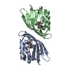
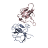
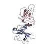
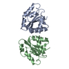
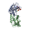
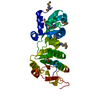
- PDB-5x3d: Crystal structure of HEP-CMP-bound form of cytidylyltransferase (... -
+ Open data

- Basic information
| Entry | Database: PDB / ID: 5x3d | ||||||
|---|---|---|---|---|---|---|---|
| Title | Crystal structure of HEP-CMP-bound form of cytidylyltransferase (CyTase) domain of Fom1 from Streptomyces wedmorensis | ||||||
 Components Components | Phosphoenolpyruvate phosphomutase | ||||||
 Keywords Keywords | TRANSFERASE / Cytidylyltransferase / Nucleotidyltransferase / fosfomycin biosynthesis | ||||||
| Function / homology |  Function and homology information Function and homology information2-hydroxyethylphosphonate cytidylyltransferase / phosphoenolpyruvate mutase / phosphoenolpyruvate mutase activity / antibiotic biosynthetic process / nucleotidyltransferase activity Similarity search - Function | ||||||
| Biological species |  Streptomyces wedmorensis (bacteria) Streptomyces wedmorensis (bacteria) | ||||||
| Method |  X-RAY DIFFRACTION / SYNCHROTRON / MAD / Resolution: 1.93 Å X-RAY DIFFRACTION / SYNCHROTRON / MAD / Resolution: 1.93 Å | ||||||
 Authors Authors | Tomita, T. / Cho, S.H. / Kuzuyama, T. / Nishiyama, M. | ||||||
 Citation Citation | Journal: ACS Chem. Biol. / Year: 2017 Title: Fosfomycin Biosynthesis via Transient Cytidylylation of 2-Hydroxyethylphosphonate by the Bifunctional Fom1 Enzyme Authors: Cho, S.H. / Kim, S.Y. / Tomita, T. / Shiraishi, T. / Park, J.S. / Sato, S. / Kudo, F. / Eguchi, T. / Funa, N. / Nishiyama, M. / Kuzuyama, T. | ||||||
| History |
|
- Structure visualization
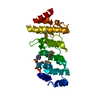
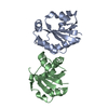
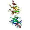
| Structure viewer | Molecule: MolmilJmol/JSmol |
|---|
- Downloads & links
-Download
| PDBx/mmCIF format | 5x3d.cif.gz | 42.3 KB | Display | PDBx/mmCIF format |
|---|---|---|---|---|
| PDB format | pdb5x3d.ent.gz | 27.8 KB | Display | PDB format |
| PDBx/mmJSON format | 5x3d.json.gz | Tree view | PDBx/mmJSON format | |
| Others |  Other downloads Other downloads |
-Validation report
| Arichive directory | https://data.pdbj.org/pub/pdb/validation_reports/x3/5x3dftp://data.pdbj.org/pub/pdb/validation_reports/x3/5x3d | HTTPS FTP |
|---|
-Related structure data
| Similar structure data |
|---|

-Links
 PDBj
PDBj
- Assembly
| Deposited unit | 
| ||||||||
|---|---|---|---|---|---|---|---|---|---|
| 1 | 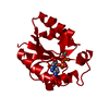
| ||||||||
| Unit cell |
|
-Components
| #1: Protein | Mass: 17817.314 Da / Num. of mol.: 1 / Fragment: Cytidylyltransferase domain, UNP residues 1-139 Source method: isolated from a genetically manipulated source Source: (gene. exp.) Streptomyces wedmorensis (bacteria) / Gene: Fom1(N) / Plasmid: pHis8 / Production host: |
|---|---|
| #2: Chemical | ChemComp-7XL / [[(  Mass: 431.230 Da / Num. of mol.: 1 / Source method: obtained synthetically / Formula: C11H19N3O11P2 Mass: 431.230 Da / Num. of mol.: 1 / Source method: obtained synthetically / Formula: C11H19N3O11P2 |
| #3: Water | ChemComp-HOH /  Mass: 18.015 Da / Num. of mol.: 73 / Source method: isolated from a natural source / Formula: H2O Mass: 18.015 Da / Num. of mol.: 73 / Source method: isolated from a natural source / Formula: H2O |
-Experimental details
-Experiment
| Experiment | Method: X-RAY DIFFRACTION / Number of used crystals: 1 |
|---|
- Sample preparation
| Crystal | Density Matthews: 2.29 Å3/Da / Density % sol: 46.37 % |
|---|---|
| Crystal grow | Temperature: 293 K / Method: vapor diffusion, hanging drop / pH: 7.5 / Details: 0.1 M HEPES, 4.3 M sodium chloride |
-Data collection
| Diffraction | Mean temperature: 95 K | ||||||||||||
|---|---|---|---|---|---|---|---|---|---|---|---|---|---|
| Diffraction source | Source: SYNCHROTRON / Site: Photon Factory  / Beamline: AR-NW12A / Wavelength: 0.97908, 0.97927, 0.96404 / Beamline: AR-NW12A / Wavelength: 0.97908, 0.97927, 0.96404 | ||||||||||||
| Detector | Type: ADSC QUANTUM 270 / Detector: CCD / Date: Nov 24, 2013 | ||||||||||||
| Radiation | Protocol: MAD / Monochromatic (M) / Laue (L): M / Scattering type: x-ray | ||||||||||||
| Radiation wavelength |
| ||||||||||||
| Reflection | Resolution: 1.93→50 Å / Num. obs: 13233 / % possible obs: 99.9 % / Redundancy: 20.9 % / Net I/σ(I): 63.1 |
- Processing
| Software |
| ||||||||||||||||||||||||||||||||||||||||||||||||||||||||||||||||||||||||||||||||||||||||||||||||||||||||||||||||||||||||||||||||||||||||||||||||||||||||||||||||||||||||||||||||||||||
|---|---|---|---|---|---|---|---|---|---|---|---|---|---|---|---|---|---|---|---|---|---|---|---|---|---|---|---|---|---|---|---|---|---|---|---|---|---|---|---|---|---|---|---|---|---|---|---|---|---|---|---|---|---|---|---|---|---|---|---|---|---|---|---|---|---|---|---|---|---|---|---|---|---|---|---|---|---|---|---|---|---|---|---|---|---|---|---|---|---|---|---|---|---|---|---|---|---|---|---|---|---|---|---|---|---|---|---|---|---|---|---|---|---|---|---|---|---|---|---|---|---|---|---|---|---|---|---|---|---|---|---|---|---|---|---|---|---|---|---|---|---|---|---|---|---|---|---|---|---|---|---|---|---|---|---|---|---|---|---|---|---|---|---|---|---|---|---|---|---|---|---|---|---|---|---|---|---|---|---|---|---|---|---|
| Refinement | Method to determine structure: MAD / Resolution: 1.93→30.05 Å / Cor.coef. Fo:Fc: 0.961 / Cor.coef. Fo:Fc free: 0.941 / SU B: 2.993 / SU ML: 0.088 / Cross valid method: THROUGHOUT / ESU R: 0.137 / ESU R Free: 0.136 / Details: HYDROGENS HAVE BEEN ADDED IN THE RIDING POSITIONS
| ||||||||||||||||||||||||||||||||||||||||||||||||||||||||||||||||||||||||||||||||||||||||||||||||||||||||||||||||||||||||||||||||||||||||||||||||||||||||||||||||||||||||||||||||||||||
| Solvent computation | Ion probe radii: 0.8 Å / Shrinkage radii: 0.8 Å / VDW probe radii: 1.2 Å | ||||||||||||||||||||||||||||||||||||||||||||||||||||||||||||||||||||||||||||||||||||||||||||||||||||||||||||||||||||||||||||||||||||||||||||||||||||||||||||||||||||||||||||||||||||||
| Displacement parameters | Biso mean: 34.472 Å2
| ||||||||||||||||||||||||||||||||||||||||||||||||||||||||||||||||||||||||||||||||||||||||||||||||||||||||||||||||||||||||||||||||||||||||||||||||||||||||||||||||||||||||||||||||||||||
| Refinement step | Cycle: 1 / Resolution: 1.93→30.05 Å
| ||||||||||||||||||||||||||||||||||||||||||||||||||||||||||||||||||||||||||||||||||||||||||||||||||||||||||||||||||||||||||||||||||||||||||||||||||||||||||||||||||||||||||||||||||||||
| Refine LS restraints |
|