 Movie
Movie Controller

[English] 日本語
 Yorodumi
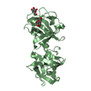
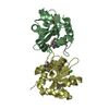
- PDB-2b7l: Crystal Structure of CTP:Glycerol-3-Phosphate Cytidylyltransferas... -
+ Open data

- Basic information
| Entry | Database: PDB / ID: 2b7l | ||||||
|---|---|---|---|---|---|---|---|
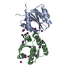
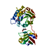
| Title | Crystal Structure of CTP:Glycerol-3-Phosphate Cytidylyltransferase from Staphylococcus aureus | ||||||
 Components Components | glycerol-3-phosphate cytidylyltransferase | ||||||
 Keywords Keywords | TRANSFERASE / cytidylyltransferase / Rossmann fold | ||||||
| Function / homology |  Function and homology information Function and homology informationglycerol-3-phosphate cytidylyltransferase / glycerol-3-phosphate cytidylyltransferase activity / teichoic acid biosynthetic process / metal ion binding / cytoplasm Similarity search - Function | ||||||
| Biological species |   Staphylococcus aureus (bacteria) Staphylococcus aureus (bacteria) | ||||||
| Method |  X-RAY DIFFRACTION / SYNCHROTRON / MOLECULAR REPLACEMENT / Resolution: 3 Å X-RAY DIFFRACTION / SYNCHROTRON / MOLECULAR REPLACEMENT / Resolution: 3 Å | ||||||
 Authors Authors | Fong, D.H. / Yim, V.C.-N. / D'Elia, M.A. / Brown, E.D. / Berghuis, A.M. | ||||||
 Citation Citation | Journal: Biochim.Biophys.Acta / Year: 2006 Title: Crystal structure of CTP:glycerol-3-phosphate cytidylyltransferase from Staphylococcus aureus: examination of structural basis for kinetic mechanism. Authors: Fong, D.H. / Yim, V.C.-N. / D'Elia, M.A. / Brown, E.D. / Berghuis, A.M. | ||||||
| History |
|
- Structure visualization
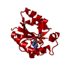
| Structure viewer | Molecule: MolmilJmol/JSmol |
|---|
- Downloads & links
-Download
| PDBx/mmCIF format | 2b7l.cif.gz | 104.8 KB | Display | PDBx/mmCIF format |
|---|---|---|---|---|
| PDB format | pdb2b7l.ent.gz | 81.8 KB | Display | PDB format |
| PDBx/mmJSON format | 2b7l.json.gz | Tree view | PDBx/mmJSON format | |
| Others |  Other downloads Other downloads |
-Validation report
| Arichive directory | https://data.pdbj.org/pub/pdb/validation_reports/b7/2b7lftp://data.pdbj.org/pub/pdb/validation_reports/b7/2b7l | HTTPS FTP |
|---|
-Related structure data
| Related structure data | 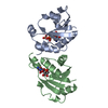 1cozS S: Starting model for refinement |
|---|---|
| Similar structure data |


-Links
 PDBj
PDBj- Assembly
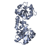
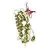
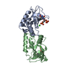
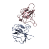
| Deposited unit | 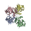
| ||||||||
|---|---|---|---|---|---|---|---|---|---|
| 1 | 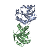
| ||||||||
| 2 | 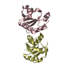
| ||||||||
| Unit cell |
|
-Components
| #1: Protein | Mass: 15843.130 Da / Num. of mol.: 4 Source method: isolated from a genetically manipulated source Source: (gene. exp.) Staphylococcus aureus (bacteria) / Plasmid: pKK223 / Species (production host): Escherichia coliProduction host: Strain (production host): W3110 References: UniProt: O05155, glycerol-3-phosphate cytidylyltransferase |
|---|
-Experimental details
-Experiment
| Experiment | Method: X-RAY DIFFRACTION / Number of used crystals: 1 |
|---|
- Sample preparation
| Crystal | Density Matthews: 2.99 Å3/Da / Density % sol: 58.9 % |
|---|---|
| Crystal grow | Temperature: 295 K / Method: vapor diffusion, hanging drop / pH: 8 Details: 8% ethylene glycol, 3% D(+) glucose, VAPOR DIFFUSION, HANGING DROP, temperature 295K |
-Data collection
| Diffraction | Mean temperature: 100 K |
|---|---|
| Diffraction source | Source: SYNCHROTRON / Site: NSLS  / Beamline: X8C / Wavelength: 1.072 Å / Beamline: X8C / Wavelength: 1.072 Å |
| Detector | Type: ADSC QUANTUM 4 / Detector: CCD / Date: Apr 27, 2000 / Details: mirror |
| Radiation | Monochromator: Si (111) double-crystal monochromator / Protocol: SINGLE WAVELENGTH / Monochromatic (M) / Laue (L): M / Scattering type: x-ray |
| Radiation wavelength | Wavelength: 1.072 Å / Relative weight: 1 |
| Reflection | Resolution: 3→50 Å / Num. all: 15217 / Num. obs: 15217 / % possible obs: 95.2 % / Observed criterion σ(F): 0 / Observed criterion σ(I): 0 / Redundancy: 7.5 % / Rmerge(I) obs: 0.135 / Rsym value: 0.135 / Net I/σ(I): 7 |
| Reflection shell | Resolution: 3→3.11 Å / % possible all: 70.2 |
- Processing
| Software |
| ||||||||||||||||||||||||||||||||||||
|---|---|---|---|---|---|---|---|---|---|---|---|---|---|---|---|---|---|---|---|---|---|---|---|---|---|---|---|---|---|---|---|---|---|---|---|---|---|
| Refinement | Method to determine structure: MOLECULAR REPLACEMENT Starting model: PDB entry 1COZ Resolution: 3→46.08 Å / Rfactor Rfree error: 0.007 / Data cutoff high absF: 179059.88 / Data cutoff low absF: 0 / Isotropic thermal model: RESTRAINED / Cross valid method: THROUGHOUT / σ(F): 0 / Stereochemistry target values: Engh & Huber
| ||||||||||||||||||||||||||||||||||||
| Solvent computation | Solvent model: FLAT MODEL / Bsol: 40.4364 Å2 / ksol: 0.404294 e/Å3 | ||||||||||||||||||||||||||||||||||||
| Displacement parameters | Biso mean: 30.6 Å2
| ||||||||||||||||||||||||||||||||||||
| Refine analyze |
| ||||||||||||||||||||||||||||||||||||
| Refinement step | Cycle: LAST / Resolution: 3→46.08 Å
| ||||||||||||||||||||||||||||||||||||
| Refine LS restraints |
| ||||||||||||||||||||||||||||||||||||
| Refine LS restraints NCS | NCS model details: CONSTR | ||||||||||||||||||||||||||||||||||||
| LS refinement shell | Resolution: 3→3.19 Å / Rfactor Rfree error: 0.03 / Total num. of bins used: 6
| ||||||||||||||||||||||||||||||||||||
| Xplor file |
|
