 Movie
Movie Controller

[English] 日本語
 Yorodumi
- PDB-5wqw: X-ray structure of catalytic domain of autolysin from Clostridium... -
+ Open data

- Basic information
| Entry | Database: PDB / ID: 5wqw | |||||||||
|---|---|---|---|---|---|---|---|---|---|---|
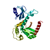
| Title | X-ray structure of catalytic domain of autolysin from Clostridium perfringens | |||||||||
 Components Components | N-acetylglucosaminidase | |||||||||
 Keywords Keywords | HYDROLASE / autolysin / glycoside hydrolase 73 family | |||||||||
| Function / homology |  Function and homology information Function and homology information | |||||||||
| Biological species |   Clostridium perfringens (bacteria) Clostridium perfringens (bacteria) | |||||||||
| Method |  X-RAY DIFFRACTION / Resolution: 1.76 Å X-RAY DIFFRACTION / Resolution: 1.76 Å | |||||||||
 Authors Authors | Tamai, E. / Sekiya, H. / Goda, E. / Makihata, N. / Maki, J. / Yoshida, H. / Kamitori, S. | |||||||||
| Funding support |  Japan, 1items Japan, 1items
| |||||||||
 Citation Citation | Journal: FEBS Lett. / Year: 2017 Title: Structural and biochemical characterization of the Clostridium perfringens autolysin catalytic domain Authors: Tamai, E. / Sekiya, H. / Goda, E. / Makihata, N. / Maki, J. / Yoshida, H. / Kamitori, S. | |||||||||
| History |
|
- Structure visualization
| Structure viewer | Molecule: MolmilJmol/JSmol |
|---|
- Downloads & links
-Download
| PDBx/mmCIF format | 5wqw.cif.gz | 67.4 KB | Display | PDBx/mmCIF format |
|---|---|---|---|---|
| PDB format | pdb5wqw.ent.gz | 48.2 KB | Display | PDB format |
| PDBx/mmJSON format | 5wqw.json.gz | Tree view | PDBx/mmJSON format | |
| Others |  Other downloads Other downloads |
-Validation report
| Summary document | 5wqw_validation.pdf.gz | 443.8 KB | Display | wwPDB validaton report |
|---|---|---|---|---|
| Full document | 5wqw_full_validation.pdf.gz | 445.8 KB | Display | |
| Data in XML | 5wqw_validation.xml.gz | 12.9 KB | Display | |
| Data in CIF | 5wqw_validation.cif.gz | 17.6 KB | Display | |
| Arichive directory | https://data.pdbj.org/pub/pdb/validation_reports/wq/5wqwftp://data.pdbj.org/pub/pdb/validation_reports/wq/5wqw | HTTPS FTP |
-Related structure data
| Similar structure data |
|---|


-Links
 PDBj
PDBj- Assembly
| Deposited unit | 
| ||||||||
|---|---|---|---|---|---|---|---|---|---|
| 1 |
| ||||||||
| Unit cell |
|
-Components
| #1: Protein | Mass: 30576.330 Da / Num. of mol.: 1 / Fragment: autolysin catalytic domain, UNP residues 871-1129 Source method: isolated from a genetically manipulated source Source: (gene. exp.) Clostridium perfringens (strain 13 / Type A) (bacteria)Strain: 13 / Type A / Gene: CPE1231 / Production host: | ||
|---|---|---|---|
| #2: Chemical | ChemComp-EDO /   Mass: 62.068 Da / Num. of mol.: 6 / Source method: obtained synthetically / Formula: C2H6O2 Mass: 62.068 Da / Num. of mol.: 6 / Source method: obtained synthetically / Formula: C2H6O2#3: Water | ChemComp-HOH / |  Mass: 18.015 Da / Num. of mol.: 121 / Source method: isolated from a natural source / Formula: H2O Mass: 18.015 Da / Num. of mol.: 121 / Source method: isolated from a natural source / Formula: H2O |
-Experimental details
-Experiment
| Experiment | Method: X-RAY DIFFRACTION / Number of used crystals: 1 |
|---|
- Sample preparation
| Crystal | Density Matthews: 2.06 Å3/Da / Density % sol: 35.81 % |
|---|---|
| Crystal grow | Temperature: 298 K / Method: vapor diffusion, sitting drop / pH: 9.5 / Details: 0.1M CHES PH9.5, 10% (W/V) PEG 3000 |
-Data collection
| Diffraction | Mean temperature: 100 K |
|---|---|
| Diffraction source | Source: ROTATING ANODE / Type: RIGAKU MICROMAX-007 HF / Wavelength: 1.5418 Å |
| Detector | Type: RIGAKU RAXIS IV / Detector: IMAGE PLATE / Date: Aug 15, 2016 |
| Radiation | Protocol: SINGLE WAVELENGTH / Monochromatic (M) / Laue (L): M / Scattering type: x-ray |
| Radiation wavelength | Wavelength: 1.5418 Å / Relative weight: 1 |
| Reflection | Resolution: 1.76→42.87 Å / Num. obs: 22481 / % possible obs: 96 % / Redundancy: 3.68 % / CC1/2: 0.998 / Rmerge(I) obs: 0.07 / Net I/σ(I): 13.3 |
| Reflection shell | Resolution: 1.76→1.82 Å / Redundancy: 3.57 % / Rmerge(I) obs: 0.42 / Mean I/σ(I) obs: 3 / CC1/2: 0.852 / % possible all: 91.4 |
- Processing
| Software |
| ||||||||||||||||||||||||||||||||||||||||||||||||||||||||||||||||||||||||||||||||||||||||||||||||||||||||||||||||||||||||||||||||||||||||||||||||||||||||||||||||||||||||||||||||||||||
|---|---|---|---|---|---|---|---|---|---|---|---|---|---|---|---|---|---|---|---|---|---|---|---|---|---|---|---|---|---|---|---|---|---|---|---|---|---|---|---|---|---|---|---|---|---|---|---|---|---|---|---|---|---|---|---|---|---|---|---|---|---|---|---|---|---|---|---|---|---|---|---|---|---|---|---|---|---|---|---|---|---|---|---|---|---|---|---|---|---|---|---|---|---|---|---|---|---|---|---|---|---|---|---|---|---|---|---|---|---|---|---|---|---|---|---|---|---|---|---|---|---|---|---|---|---|---|---|---|---|---|---|---|---|---|---|---|---|---|---|---|---|---|---|---|---|---|---|---|---|---|---|---|---|---|---|---|---|---|---|---|---|---|---|---|---|---|---|---|---|---|---|---|---|---|---|---|---|---|---|---|---|---|---|
| Refinement | Resolution: 1.76→42.87 Å / Cor.coef. Fo:Fc: 0.96 / Cor.coef. Fo:Fc free: 0.944 / SU B: 2.09 / SU ML: 0.067 / Cross valid method: THROUGHOUT / ESU R: 0.12 / ESU R Free: 0.111
| ||||||||||||||||||||||||||||||||||||||||||||||||||||||||||||||||||||||||||||||||||||||||||||||||||||||||||||||||||||||||||||||||||||||||||||||||||||||||||||||||||||||||||||||||||||||
| Solvent computation | Ion probe radii: 0.8 Å / Shrinkage radii: 0.8 Å / VDW probe radii: 1.2 Å | ||||||||||||||||||||||||||||||||||||||||||||||||||||||||||||||||||||||||||||||||||||||||||||||||||||||||||||||||||||||||||||||||||||||||||||||||||||||||||||||||||||||||||||||||||||||
| Displacement parameters | Biso mean: 17.89 Å2
| ||||||||||||||||||||||||||||||||||||||||||||||||||||||||||||||||||||||||||||||||||||||||||||||||||||||||||||||||||||||||||||||||||||||||||||||||||||||||||||||||||||||||||||||||||||||
| Refinement step | Cycle: 1 / Resolution: 1.76→42.87 Å
| ||||||||||||||||||||||||||||||||||||||||||||||||||||||||||||||||||||||||||||||||||||||||||||||||||||||||||||||||||||||||||||||||||||||||||||||||||||||||||||||||||||||||||||||||||||||
| Refine LS restraints |
|