 Movie
Movie Controller

[English] 日本語
 Yorodumi
- PDB-5wp9: Structural Basis of Mitochondrial Receptor Binding and Constricti... -
+ Open data

- Basic information
| Entry | Database: PDB / ID: 5wp9 | |||||||||||||||
|---|---|---|---|---|---|---|---|---|---|---|---|---|---|---|---|---|
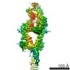
| Title | Structural Basis of Mitochondrial Receptor Binding and Constriction by Dynamin-Related Protein 1 | |||||||||||||||
 Components Components |
| |||||||||||||||
 Keywords Keywords | PROTEIN FIBRIL / Mitochondrial division / Dynamin related-protein-1 / Nucleotide / MID49 | |||||||||||||||
| Function / homology |  Function and homology information Function and homology informationmitochondrial membrane fission / regulation of ATP metabolic process / regulation of peroxisome organization / mitocytosis / Apoptotic execution phase / dynamin GTPase / regulation of mitophagy / peroxisome fission / mitochondrial fragmentation involved in apoptotic process / GTP-dependent protein binding ...mitochondrial membrane fission / regulation of ATP metabolic process / regulation of peroxisome organization / mitocytosis / Apoptotic execution phase / dynamin GTPase / regulation of mitophagy / peroxisome fission / mitochondrial fragmentation involved in apoptotic process / GTP-dependent protein binding / protein localization to mitochondrion / mitochondrial fission / regulation of mitochondrion organization / positive regulation of neutrophil chemotaxis / positive regulation of mitochondrial fission / heart contraction / intracellular distribution of mitochondria / protein complex oligomerization / brush border / positive regulation of protein targeting to membrane / clathrin-coated pit / GTPase activator activity / positive regulation of protein secretion / mitochondrion organization / small GTPase binding / endocytosis / calcium ion transport / rhythmic process / peroxisome / synaptic vesicle membrane / regulation of gene expression / microtubule binding / microtubule / mitochondrial outer membrane / GTPase activity / ubiquitin protein ligase binding / lipid binding / endoplasmic reticulum membrane / GTP binding / perinuclear region of cytoplasm / Golgi apparatus / endoplasmic reticulum / protein homodimerization activity / protein-containing complex / mitochondrion / membrane / identical protein binding / cytoplasm / cytosol Similarity search - Function | |||||||||||||||
| Biological species |  Homo sapiens (human) Homo sapiens (human) | |||||||||||||||
| Method | ELECTRON MICROSCOPY / helical reconstruction / cryo EM / Resolution: 4.22 Å | |||||||||||||||
 Authors Authors | Kalia, R. / Wang, R.Y.R. / Yusuf, A. / Thomas, P.V. / Agard, D.A. / Shaw, J.M. / Frost, A. | |||||||||||||||
| Funding support |  United States, 4items United States, 4items
| |||||||||||||||
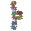
 Citation Citation | Journal: Nature / Year: 2018 Title: Structural basis of mitochondrial receptor binding and constriction by DRP1. Authors: Raghav Kalia / Ray Yu-Ruei Wang / Ali Yusuf / Paul V Thomas / David A Agard / Janet M Shaw / Adam Frost / Abstract: Mitochondrial inheritance, genome maintenance and metabolic adaptation depend on organelle fission by dynamin-related protein 1 (DRP1) and its mitochondrial receptors. DRP1 receptors include the ...Mitochondrial inheritance, genome maintenance and metabolic adaptation depend on organelle fission by dynamin-related protein 1 (DRP1) and its mitochondrial receptors. DRP1 receptors include the paralogues mitochondrial dynamics proteins of 49 and 51 kDa (MID49 and MID51) and mitochondrial fission factor (MFF); however, the mechanisms by which these proteins recruit and regulate DRP1 are unknown. Here we present a cryo-electron microscopy structure of full-length human DRP1 co-assembled with MID49 and an analysis of structure- and disease-based mutations. We report that GTP induces a marked elongation and rotation of the GTPase domain, bundle-signalling element and connecting hinge loops of DRP1. In this conformation, a network of multivalent interactions promotes the polymerization of a linear DRP1 filament with MID49 or MID51. After co-assembly, GTP hydrolysis and exchange lead to MID receptor dissociation, filament shortening and curling of DRP1 oligomers into constricted and closed rings. Together, these views of full-length, receptor- and nucleotide-bound conformations reveal how DRP1 performs mechanical work through nucleotide-driven allostery. | |||||||||||||||
| History |
|
- Structure visualization
| Movie |
Movie viewer |
|---|---|
| Structure viewer | Molecule: MolmilJmol/JSmol |
- Downloads & links
-Download
| PDBx/mmCIF format | 5wp9.cif.gz | 2.6 MB | Display | PDBx/mmCIF format |
|---|---|---|---|---|
| PDB format | pdb5wp9.ent.gz | Display | PDB format | |
| PDBx/mmJSON format | 5wp9.json.gz | Tree view | PDBx/mmJSON format | |
| Others |  Other downloads Other downloads |
-Validation report
| Arichive directory | https://data.pdbj.org/pub/pdb/validation_reports/wp/5wp9ftp://data.pdbj.org/pub/pdb/validation_reports/wp/5wp9 | HTTPS FTP |
|---|
-Related structure data
| Related structure data |  8874MC M: map data used to model this data C: citing same article ( |
|---|---|
| Similar structure data |


-Links
 PDBj
PDBj- Assembly
| Deposited unit | 
|
|---|---|
| 1 |
|
| Symmetry | Helical symmetry: (Circular symmetry: 1 / Dyad axis: no / N subunits divisor: 1 / Num. of operations: 20 / Rise per n subunits: 54.8 Å / Rotation per n subunits: -0.8 °) |
-Components
| #1: Protein | Mass: 79546.453 Da / Num. of mol.: 8 Source method: isolated from a genetically manipulated source Source: (gene. exp.) Homo sapiens (human) / Gene: DNM1L, DLP1, DRP1 / Production host:  #2: Protein | Mass: 36156.570 Da / Num. of mol.: 8 / Fragment: UNP residues 126-454 Source method: isolated from a genetically manipulated source Source: (gene. exp.) Homo sapiens (human) / Gene: MIEF2, MID49, SMCR7 / Production host: #3: Chemical | ChemComp-GCP /   Mass: 521.208 Da / Num. of mol.: 8 / Source method: obtained synthetically / Formula: C11H18N5O13P3 / Comment: GMP-PCP, energy-carrying molecule analogue*YM Mass: 521.208 Da / Num. of mol.: 8 / Source method: obtained synthetically / Formula: C11H18N5O13P3 / Comment: GMP-PCP, energy-carrying molecule analogue*YM#4: Chemical | ChemComp-MG /   Mass: 24.305 Da / Num. of mol.: 8 / Source method: obtained synthetically / Formula: Mg Mass: 24.305 Da / Num. of mol.: 8 / Source method: obtained synthetically / Formula: Mg |
|---|
-Experimental details
-Experiment
| Experiment | Method: ELECTRON MICROSCOPY |
|---|---|
| EM experiment | Aggregation state: FILAMENT / 3D reconstruction method: helical reconstruction |
- Sample preparation
| Component | Name: DRP1-MID49 / Type: COMPLEX Details: CryoEM structure of Dynamin-Related Protein 1 in complex with Adaptor MID49 Entity ID: #1-#2 / Source: RECOMBINANT |
|---|---|
| Molecular weight | Value: 115.56 kDa/nm / Experimental value: YES |
| Source (natural) | Organism: Homo sapiens (human) |
| Source (recombinant) | Organism: |
| Buffer solution | pH: 7.5 |
| Specimen | Embedding applied: NO / Shadowing applied: NO / Staining applied: NO / Vitrification applied: YES |
| Specimen support | Grid material: COPPER / Grid mesh size: 200 divisions/in. / Grid type: Quantifoil R2/2 |
| Vitrification | Cryogen name: ETHANE |
- Electron microscopy imaging
| Experimental equipment |  Model: Tecnai F30 / Image courtesy: FEI Company |
|---|---|
| Microscopy | Model: FEI TECNAI F30 |
| Electron gun | Electron source:  FIELD EMISSION GUN / Accelerating voltage: 300 kV / Illumination mode: FLOOD BEAM FIELD EMISSION GUN / Accelerating voltage: 300 kV / Illumination mode: FLOOD BEAM |
| Electron lens | Mode: BRIGHT FIELD / Nominal magnification: 31000 X / Nominal defocus max: 4000 nm / Nominal defocus min: 300 nm / Cs: 2 mm / C2 aperture diameter: 30 µm |
| Image recording | Electron dose: 60 e/Å2 / Detector mode: SUPER-RESOLUTION / Film or detector model: GATAN K2 SUMMIT (4k x 4k) |
- Processing
| EM software | Name: SerialEM / Category: image acquisition |
|---|---|
| CTF correction | Type: NONE |
| Helical symmerty | Angular rotation/subunit: -0.8 ° / Axial rise/subunit: 54.8 Å / Axial symmetry: C1 |
| 3D reconstruction | Resolution: 4.22 Å / Resolution method: FSC 0.143 CUT-OFF / Num. of particles: 412684 / Symmetry type: HELICAL |
