[English] 日本語
Yorodumi
- PDB-5wnj: Crystal structure of murine receptor-interacting protein kinase 4... -

+
Open data


ID or keywords:

Loading...

-
Basic information

Entry
Database: PDB / ID: 5wnj
TitleCrystal structure of murine receptor-interacting protein kinase 4 (Ripk4) D143N in complex with lestaurtinib
ComponentsReceptor-interacting serine/threonine-protein kinase 4
KeywordsTRANSFERASE/TRANSFERASE INHIBITOR / Kinase / Inhibitor / Complex / TRANSFERASE / TRANSFERASE-TRANSFERASE INHIBITOR complex
Function / homology
Function and homology information


morphogenesis of an epithelium / non-specific serine/threonine protein kinase / phosphorylation / protein serine kinase activity / protein serine/threonine kinase activity / ATP binding / membrane / cytoplasm
Similarity search - Function
Ankyrin repeats (many copies) / Ankyrin repeats (3 copies) / Ankyrin repeat profile. / Ankyrin repeat region circular profile. / ankyrin repeats / Ankyrin repeat / Ankyrin repeat-containing domain superfamily / Transferase(Phosphotransferase) domain 1 / Transferase(Phosphotransferase); domain 1 / Serine/threonine-protein kinase, active site ...Ankyrin repeats (many copies) / Ankyrin repeats (3 copies) / Ankyrin repeat profile. / Ankyrin repeat region circular profile. / ankyrin repeats / Ankyrin repeat / Ankyrin repeat-containing domain superfamily / Transferase(Phosphotransferase) domain 1 / Transferase(Phosphotransferase); domain 1 / Serine/threonine-protein kinase, active site / Serine/Threonine protein kinases active-site signature. / Protein kinase domain / Serine/Threonine protein kinases, catalytic domain / Protein kinase, ATP binding site / Protein kinases ATP-binding region signature. / Protein kinase domain profile. / Protein kinase domain / Protein kinase-like domain superfamily / Orthogonal Bundle / Mainly Alpha
Similarity search - Domain/homology
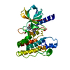
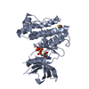
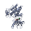
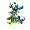
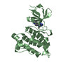
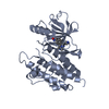
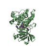
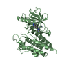
Lestaurtinib / Receptor-interacting serine/threonine-protein kinase 4
Similarity search - Component
Biological speciesMus musculus (house mouse)
MethodX-RAY DIFFRACTION / SYNCHROTRON / MOLECULAR REPLACEMENT / Resolution: 2.55 Å
AuthorsHuang, C.S. / Hymowitz, S.G.
CitationJournal: Structure / Year: 2018
Title: Crystal Structure of Ripk4 Reveals Dimerization-Dependent Kinase Activity.
Authors: Huang, C.S. / Oberbeck, N. / Hsiao, Y.C. / Liu, P. / Johnson, A.R. / Dixit, V.M. / Hymowitz, S.G.
History
DepositionAug 1, 2017Deposition site: RCSB / Processing site: RCSB
Revision 1.0May 9, 2018Provider: repository / Type: Initial release
Revision 1.1May 16, 2018Group: Data collection / Database references / Category: citation / citation_author
Item: _citation.journal_id_ISSN / _citation.journal_volume ..._citation.journal_id_ISSN / _citation.journal_volume / _citation.page_first / _citation.pdbx_database_id_PubMed / _citation.title / _citation_author.name
Revision 2.0Dec 7, 2022Group: Database references / Non-polymer description / Structure summary
Category: chem_comp / database_2 / entity
Item: _chem_comp.formula / _chem_comp.formula_weight ..._chem_comp.formula / _chem_comp.formula_weight / _database_2.pdbx_DOI / _database_2.pdbx_database_accession / _entity.formula_weight

-
Structure visualization

Structure viewerMolecule:
MolmilJmol/JSmol

Downloads & links

-
Assembly

Deposited unit
A: Receptor-interacting serine/threonine-protein kinase 4
hetero molecules


Theoretical massNumber of molelcules
Total (without water)39,4633
Polymers38,9881
Non-polymers4752
Water43224
1


  • Idetical with deposited unit
  • defined by author
  • Evidence: mass spectrometry
TypeNameSymmetry operationNumber
identity operation1_555x,y,z1
2
A: Receptor-interacting serine/threonine-protein kinase 4
hetero molecules

A: Receptor-interacting serine/threonine-protein kinase 4
hetero molecules


Theoretical massNumber of molelcules
Total (without water)78,9266
Polymers77,9762
Non-polymers9504
Water362
TypeNameSymmetry operationNumber
identity operation1_555x,y,z1
crystal symmetry operation8_554x,-y,-z-1/21
Buried area2970 Å2
ΔGint-28 kcal/mol
Surface area23440 Å2
MethodPISA
Unit cell
Length a, b, c (Å)70.825, 110.247, 145.536
Angle α, β, γ (deg.)90.00, 90.00, 90.00
Int Tables number24
Space group name H-MI212121

-
Components

#1: Protein Receptor-interacting serine/threonine-protein kinase 4 / Ankyrin repeat domain-containing protein 3 / PKC-associated protein kinase / PKC-regulated protein kinase


Mass: 38988.039 Da / Num. of mol.: 1 / Fragment: residues 1-342 / Mutation: D143N
Source method: isolated from a genetically manipulated source
Source: (gene. exp.) Mus musculus (house mouse) / Gene: Ripk4, Ankrd3, Pkk / Production host: 
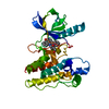
Spodoptera frugiperda (fall armyworm)
References: UniProt: Q9ERK0, non-specific serine/threonine protein kinase
#2: Chemical ChemComp-2V9 / Lestaurtinib / (5S,6S,8R)-6-hydroxy-6-(hydroxymethyl)-5-methyl-5,6,7,8-tetrahydro-13H-5,8-epoxy-4b,8a,14-triazadibenzo[b,h]cycloocta[1,2,3,4-jkl]cyclopenta[e]-as-indacen-13-one / Lestaurtinib


Mass: 439.463 Da / Num. of mol.: 1 / Source method: obtained synthetically / Formula: C26H21N3O4 / Comment: inhibitor*YM
#3: Chemical ChemComp-CL / CHLORIDE ION / Chloride


Mass: 35.453 Da / Num. of mol.: 1 / Source method: isolated from a natural source / Formula: Cl
#4: Water ChemComp-HOH / water / Water


Mass: 18.015 Da / Num. of mol.: 24 / Source method: isolated from a natural source / Formula: H2O

-
Experimental details

-
Experiment

ExperimentMethod: X-RAY DIFFRACTION / Number of used crystals: 1

-
Sample preparation

CrystalDensity Matthews: 3.64 Å3/Da / Density % sol: 66.24 %
Crystal growTemperature: 292 K / Method: microbatch
Details: 0.1M HEPES pH 7.5, 0.2M magnesium chloride, 10% 2-propanol, 15% ethylene glycol, 10% PEG4000

-
Data collection

DiffractionMean temperature: 100 K
Diffraction sourceSource: SYNCHROTRON / Site: ALS / Beamline: 5.0.2 / Wavelength: 1 Å
DetectorType: DECTRIS PILATUS 6M / Detector: PIXEL / Date: Jun 3, 2015
RadiationProtocol: SINGLE WAVELENGTH / Monochromatic (M) / Laue (L): M / Scattering type: x-ray
Radiation wavelengthWavelength: 1 Å / Relative weight: 1
ReflectionResolution: 2.55→50.01 Å / Num. obs: 18791 / % possible obs: 99.6 % / Redundancy: 6 % / Rmerge(I) obs: 0.083 / Net I/σ(I): 19.1

-
Processing

Software
NameVersionClassification
REFMAC5.8.0158refinement
HKL-2000data reduction
HKL-2000data scaling
PHASERphasing
RefinementMethod to determine structure: MOLECULAR REPLACEMENT / Resolution: 2.55→50.01 Å / Cor.coef. Fo:Fc: 0.94 / Cor.coef. Fo:Fc free: 0.918 / SU B: 7.865 / SU ML: 0.17 / Cross valid method: THROUGHOUT / ESU R: 0.269 / ESU R Free: 0.225 / Stereochemistry target values: MAXIMUM LIKELIHOOD / Details: HYDROGENS HAVE BEEN ADDED IN THE RIDING POSITIONS
RfactorNum. reflection% reflectionSelection details
Rfree0.25226 912 4.9 %RANDOM
Rwork0.2195 ---
obs0.22105 17877 99.28 %-
Solvent computationIon probe radii: 0.8 Å / Shrinkage radii: 0.8 Å / VDW probe radii: 1.2 Å / Solvent model: MASK
Displacement parametersBiso mean: 63.227 Å2
Baniso -1Baniso -2Baniso -3
1-2.98 Å20 Å20 Å2
2--1.05 Å20 Å2
3----4.03 Å2
Refinement stepCycle: 1 / Resolution: 2.55→50.01 Å
ProteinNucleic acidLigandSolventTotal
Num. atoms2042 0 34 24 2100
Refine LS restraints
Refine-IDTypeDev idealDev ideal targetNumber
X-RAY DIFFRACTIONr_bond_refined_d0.0080.0192137
X-RAY DIFFRACTIONr_bond_other_d0.0010.021942
X-RAY DIFFRACTIONr_angle_refined_deg1.2321.9582917
X-RAY DIFFRACTIONr_angle_other_deg0.9072.9884481
X-RAY DIFFRACTIONr_dihedral_angle_1_deg5.6435261
X-RAY DIFFRACTIONr_dihedral_angle_2_deg32.95822.85784
X-RAY DIFFRACTIONr_dihedral_angle_3_deg12.72115336
X-RAY DIFFRACTIONr_dihedral_angle_4_deg18.5821513
X-RAY DIFFRACTIONr_chiral_restr0.0680.2320
X-RAY DIFFRACTIONr_gen_planes_refined0.0060.0212361
X-RAY DIFFRACTIONr_gen_planes_other0.0010.02440
X-RAY DIFFRACTIONr_nbd_refined
X-RAY DIFFRACTIONr_nbd_other
X-RAY DIFFRACTIONr_nbtor_refined
X-RAY DIFFRACTIONr_nbtor_other
X-RAY DIFFRACTIONr_xyhbond_nbd_refined
X-RAY DIFFRACTIONr_xyhbond_nbd_other
X-RAY DIFFRACTIONr_metal_ion_refined
X-RAY DIFFRACTIONr_metal_ion_other
X-RAY DIFFRACTIONr_symmetry_vdw_refined
X-RAY DIFFRACTIONr_symmetry_vdw_other
X-RAY DIFFRACTIONr_symmetry_hbond_refined
X-RAY DIFFRACTIONr_symmetry_hbond_other
X-RAY DIFFRACTIONr_symmetry_metal_ion_refined
X-RAY DIFFRACTIONr_symmetry_metal_ion_other
X-RAY DIFFRACTIONr_mcbond_it3.1956.6621053
X-RAY DIFFRACTIONr_mcbond_other3.1766.6581052
X-RAY DIFFRACTIONr_mcangle_it5.1229.9671311
X-RAY DIFFRACTIONr_mcangle_other5.129.9731312
X-RAY DIFFRACTIONr_scbond_it2.9996.5791084
X-RAY DIFFRACTIONr_scbond_other2.9986.5791085
X-RAY DIFFRACTIONr_scangle_it
X-RAY DIFFRACTIONr_scangle_other4.8169.771607
X-RAY DIFFRACTIONr_long_range_B_refined7.13973.8742343
X-RAY DIFFRACTIONr_long_range_B_other7.13773.8572344
X-RAY DIFFRACTIONr_rigid_bond_restr
X-RAY DIFFRACTIONr_sphericity_free
X-RAY DIFFRACTIONr_sphericity_bonded
LS refinement shellResolution: 2.553→2.619 Å / Total num. of bins used: 20
RfactorNum. reflection% reflection
Rfree0.318 69 -
Rwork0.307 1253 -
obs--96.22 %

+
About Yorodumi

-
News

-
Feb 9, 2022. New format data for meta-information of EMDB entries

New format data for meta-information of EMDB entries

  • Version 3 of the EMDB header file is now the official format.
  • The previous official version 1.9 will be removed from the archive.

Related info.:EMDB header

External links:wwPDB to switch to version 3 of the EMDB data model

-
Aug 12, 2020. Covid-19 info

Covid-19 info

URL: https://pdbj.org/emnavi/covid19.php

New page: Covid-19 featured information page in EM Navigator.

Related info.:Covid-19 info / Mar 5, 2020. Novel coronavirus structure data

+
Mar 5, 2020. Novel coronavirus structure data

Novel coronavirus structure data

Related info.:Yorodumi Speices / Aug 12, 2020. Covid-19 info

External links:COVID-19 featured content - PDBj / Molecule of the Month (242):Coronavirus Proteases

+
Jan 31, 2019. EMDB accession codes are about to change! (news from PDBe EMDB page)

EMDB accession codes are about to change! (news from PDBe EMDB page)

  • The allocation of 4 digits for EMDB accession codes will soon come to an end. Whilst these codes will remain in use, new EMDB accession codes will include an additional digit and will expand incrementally as the available range of codes is exhausted. The current 4-digit format prefixed with “EMD-” (i.e. EMD-XXXX) will advance to a 5-digit format (i.e. EMD-XXXXX), and so on. It is currently estimated that the 4-digit codes will be depleted around Spring 2019, at which point the 5-digit format will come into force.
  • The EM Navigator/Yorodumi systems omit the EMD- prefix.

Related info.:Q: What is EMD? / ID/Accession-code notation in Yorodumi/EM Navigator

External links:EMDB Accession Codes are Changing Soon! / Contact to PDBj

+
Jul 12, 2017. Major update of PDB

Major update of PDB

  • wwPDB released updated PDB data conforming to the new PDBx/mmCIF dictionary.
  • This is a major update changing the version number from 4 to 5, and with Remediation, in which all the entries are updated.
  • In this update, many items about electron microscopy experimental information are reorganized (e.g. em_software).
  • Now, EM Navigator and Yorodumi are based on the updated data.

External links:wwPDB Remediation / Enriched Model Files Conforming to OneDep Data Standards Now Available in the PDB FTP Archive

-
Yorodumi

Thousand views of thousand structures

  • Yorodumi is a browser for structure data from EMDB, PDB, SASBDB, etc.
  • This page is also the successor to EM Navigator detail page, and also detail information page/front-end page for Omokage search.
  • The word "yorodu" (or yorozu) is an old Japanese word meaning "ten thousand". "mi" (miru) is to see.

Related info.:EMDB / PDB / SASBDB / Comparison of 3 databanks / Yorodumi Search / Aug 31, 2016. New EM Navigator & Yorodumi / Yorodumi Papers / Jmol/JSmol / Function and homology information / Changes in new EM Navigator and Yorodumi

Read more