 Movie
Movie Controller

[English] 日本語
 Yorodumi
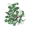
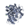
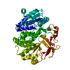
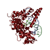
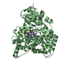
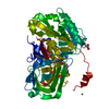
- PDB-5wjc: Crystal structure of Schizosaccharomyces pombe Mis16 in complex w... -
+ Open data

- Basic information
| Entry | Database: PDB / ID: 5wjc | |||||||||||||||
|---|---|---|---|---|---|---|---|---|---|---|---|---|---|---|---|---|
| Title | Crystal structure of Schizosaccharomyces pombe Mis16 in complex with Eic1 | |||||||||||||||
 Components Components |
| |||||||||||||||
 Keywords Keywords | PROTEIN BINDING / Fission yeast chaperone for histone H4 / Subcomponent of Mis18 complex / Eic1 binding / WD-40 repeats domain | |||||||||||||||
| Function / homology |  Function and homology information Function and homology informationNeddylation / HATs acetylate histones / RMTs methylate histone arginines / HDACs deacetylate histones / CENP-A recruiting complex / : / chromosome, centromeric core domain / H3-H4 histone complex chaperone activity / CENP-A containing chromatin assembly / attachment of mitotic spindle microtubules to kinetochore ...Neddylation / HATs acetylate histones / RMTs methylate histone arginines / HDACs deacetylate histones / CENP-A recruiting complex / : / chromosome, centromeric core domain / H3-H4 histone complex chaperone activity / CENP-A containing chromatin assembly / attachment of mitotic spindle microtubules to kinetochore / chromosome, centromeric region / kinetochore / histone binding / chromatin remodeling / cell division / nucleoplasm / nucleus / cytoplasm Similarity search - Function | |||||||||||||||
| Biological species |  | |||||||||||||||
| Method |  X-RAY DIFFRACTION / SYNCHROTRON / MOLECULAR REPLACEMENT / Resolution: 2.298 Å X-RAY DIFFRACTION / SYNCHROTRON / MOLECULAR REPLACEMENT / Resolution: 2.298 Å | |||||||||||||||
 Authors Authors | An, S. / Cho, U.-S. / Koldewey, P. / Chik, J. / Subramanian, L. | |||||||||||||||
| Funding support |  United States, 4items United States, 4items
| |||||||||||||||
 Citation Citation | Journal: Structure / Year: 2018 Title: Mis16 Switches Function from a Histone H4 Chaperone to a CENP-ACnp1-Specific Assembly Factor through Eic1 Interaction. Authors: An, S. / Koldewey, P. / Chik, J. / Subramanian, L. / Cho, U.S. | |||||||||||||||
| History |
|
- Structure visualization
| Structure viewer | Molecule: MolmilJmol/JSmol |
|---|
- Downloads & links
-Download
| PDBx/mmCIF format | 5wjc.cif.gz | 98.1 KB | Display | PDBx/mmCIF format |
|---|---|---|---|---|
| PDB format | pdb5wjc.ent.gz | 72.6 KB | Display | PDB format |
| PDBx/mmJSON format | 5wjc.json.gz | Tree view | PDBx/mmJSON format | |
| Others |  Other downloads Other downloads |
-Validation report
| Arichive directory | https://data.pdbj.org/pub/pdb/validation_reports/wj/5wjcftp://data.pdbj.org/pub/pdb/validation_reports/wj/5wjc | HTTPS FTP |
|---|
-Related structure data
| Related structure data | 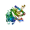 4xyhS S: Starting model for refinement |
|---|---|
| Similar structure data |


-Links
 PDBj
PDBj- Assembly
| Deposited unit | 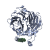
| ||||||||
|---|---|---|---|---|---|---|---|---|---|
| 1 |
| ||||||||
| Unit cell |
|
-Components
| #1: Protein | Mass: 48481.555 Da / Num. of mol.: 1 Source method: isolated from a genetically manipulated source Source: (gene. exp.) Strain: 972 / ATCC 24843 / Gene: mis16, hat2, SPCC1672.10 / Production host:  Baculovirus expression vector pFastBac1-HM / References: UniProt: O94244 Baculovirus expression vector pFastBac1-HM / References: UniProt: O94244 |
|---|---|
| #2: Protein | Mass: 13453.742 Da / Num. of mol.: 1 Source method: isolated from a genetically manipulated source Source: (gene. exp.) Strain: 972 / ATCC 24843 / Gene: SPBC27B12.02, SPBC30B4.10 / Production host: Baculovirus expression vector pFastBac1-HM / References: UniProt: O42995 |
| #3: Water | ChemComp-HOH /  Mass: 18.015 Da / Num. of mol.: 168 / Source method: isolated from a natural source / Formula: H2O Mass: 18.015 Da / Num. of mol.: 168 / Source method: isolated from a natural source / Formula: H2O |
-Experimental details
-Experiment
| Experiment | Method: X-RAY DIFFRACTION / Number of used crystals: 1 |
|---|
- Sample preparation
| Crystal | Density Matthews: 2.58 Å3/Da / Density % sol: 52.24 % / Description: Thick needle |
|---|---|
| Crystal grow | Temperature: 296.15 K / Method: vapor diffusion, hanging drop / pH: 8.5 / Details: 100 mM Tris-HCl pH 8.5, 25% PEG 2000MME |
-Data collection
| Diffraction | Mean temperature: 105 K |
|---|---|
| Diffraction source | Source: SYNCHROTRON / Site: APS / Beamline: 21-ID-G / Wavelength: 0.9786 Å |
| Detector | Type: MARMOSAIC 300 mm CCD / Detector: CCD / Date: Jun 15, 2016 |
| Radiation | Protocol: SINGLE WAVELENGTH / Monochromatic (M) / Laue (L): M / Scattering type: x-ray |
| Radiation wavelength | Wavelength: 0.9786 Å / Relative weight: 1 |
| Reflection | Resolution: 2.3→50 Å / Num. obs: 26459 / % possible obs: 90.8 % / Redundancy: 8 % / Rmerge(I) obs: 0.097 / Rpim(I) all: 0.035 / Net I/σ(I): 22.4 |
| Reflection shell | Resolution: 2.3→2.38 Å / Redundancy: 4 % / Rmerge(I) obs: 0.76 / Mean I/σ(I) obs: 1.6 / Num. unique obs: 1698 / Rpim(I) all: 0.39 / % possible all: 59.7 |
- Processing
| Software |
| |||||||||||||||||||||||||||||||||||||||||||||||||||||||||||||||||||||||||||||||||||||||||||||||||||||||||
|---|---|---|---|---|---|---|---|---|---|---|---|---|---|---|---|---|---|---|---|---|---|---|---|---|---|---|---|---|---|---|---|---|---|---|---|---|---|---|---|---|---|---|---|---|---|---|---|---|---|---|---|---|---|---|---|---|---|---|---|---|---|---|---|---|---|---|---|---|---|---|---|---|---|---|---|---|---|---|---|---|---|---|---|---|---|---|---|---|---|---|---|---|---|---|---|---|---|---|---|---|---|---|---|---|---|---|
| Refinement | Method to determine structure: MOLECULAR REPLACEMENT Starting model: 4XYH Resolution: 2.298→48.886 Å / SU ML: 0.22 / Cross valid method: FREE R-VALUE / σ(F): 1.35 / Phase error: 22.12 / Stereochemistry target values: ML
| |||||||||||||||||||||||||||||||||||||||||||||||||||||||||||||||||||||||||||||||||||||||||||||||||||||||||
| Solvent computation | Shrinkage radii: 0.9 Å / VDW probe radii: 1.11 Å / Solvent model: FLAT BULK SOLVENT MODEL | |||||||||||||||||||||||||||||||||||||||||||||||||||||||||||||||||||||||||||||||||||||||||||||||||||||||||
| Refinement step | Cycle: LAST / Resolution: 2.298→48.886 Å
| |||||||||||||||||||||||||||||||||||||||||||||||||||||||||||||||||||||||||||||||||||||||||||||||||||||||||
| Refine LS restraints |
| |||||||||||||||||||||||||||||||||||||||||||||||||||||||||||||||||||||||||||||||||||||||||||||||||||||||||
| LS refinement shell |
|
