 Movie
Movie Controller

[English] 日本語
 Yorodumi
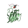
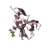
- PDB-5w7x: Crystal Structure of FHA domain of human APLF in complex with XRC... -
+ Open data

- Basic information
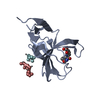
| Entry | Database: PDB / ID: 5w7x | ||||||
|---|---|---|---|---|---|---|---|
| Title | Crystal Structure of FHA domain of human APLF in complex with XRCC1 bisphospho peptide | ||||||
 Components Components |
| ||||||
 Keywords Keywords | PROTEIN BINDING / scaffold protein / DNA repair / NHEJ | ||||||
| Function / homology |  Function and homology information Function and homology informationoxidized DNA binding / telomeric DNA-containing double minutes formation / ERCC4-ERCC1 complex / negative regulation of protection from non-homologous end joining at telomere / ADP-D-ribose modification-dependent protein binding / negative regulation of protein ADP-ribosylation / regulation of isotype switching / poly-ADP-D-ribose binding / regulation of base-excision repair / histone chaperone activity ...oxidized DNA binding / telomeric DNA-containing double minutes formation / ERCC4-ERCC1 complex / negative regulation of protection from non-homologous end joining at telomere / ADP-D-ribose modification-dependent protein binding / negative regulation of protein ADP-ribosylation / regulation of isotype switching / poly-ADP-D-ribose binding / regulation of base-excision repair / histone chaperone activity / regulation of epithelial to mesenchymal transition / HDR through MMEJ (alt-NHEJ) / single strand break repair / 3' overhang single-stranded DNA endonuclease activity / response to hydroperoxide / Resolution of AP sites via the single-nucleotide replacement pathway / APEX1-Independent Resolution of AP Sites via the Single Nucleotide Replacement Pathway / DNA repair-dependent chromatin remodeling / site of DNA damage / embryo implantation / protein localization to chromatin / 3'-5' exonuclease activity / DNA-(apurinic or apyrimidinic site) endonuclease activity / protein folding chaperone / hippocampus development / Gap-filling DNA repair synthesis and ligation in GG-NER / DNA endonuclease activity / base-excision repair / double-strand break repair via nonhomologous end joining / Gap-filling DNA repair synthesis and ligation in TC-NER / double-strand break repair / site of double-strand break / histone binding / Hydrolases; Acting on ester bonds / chromosome, telomeric region / nucleotide binding / DNA repair / DNA damage response / nucleolus / chromatin / enzyme binding / zinc ion binding / nucleoplasm / nucleus / cytosol Similarity search - Function | ||||||
| Biological species |  Homo sapiens (human) Homo sapiens (human) | ||||||
| Method |  X-RAY DIFFRACTION / MOLECULAR REPLACEMENT / Resolution: 2.005 Å X-RAY DIFFRACTION / MOLECULAR REPLACEMENT / Resolution: 2.005 Å | ||||||
 Authors Authors | Pedersen, L.C. / Kim, K. / London, R.E. | ||||||
 Citation Citation | Journal: Nucleic Acids Res. / Year: 2017 Title: Characterization of the APLF FHA-XRCC1 phosphopeptide interaction and its structural and functional implications. Authors: Kim, K. / Pedersen, L.C. / Kirby, T.W. / DeRose, E.F. / London, R.E. | ||||||
| History |
|
- Structure visualization
| Structure viewer | Molecule: MolmilJmol/JSmol |
|---|
- Downloads & links
-Download
| PDBx/mmCIF format | 5w7x.cif.gz | 103.2 KB | Display | PDBx/mmCIF format |
|---|---|---|---|---|
| PDB format | pdb5w7x.ent.gz | 78.6 KB | Display | PDB format |
| PDBx/mmJSON format | 5w7x.json.gz | Tree view | PDBx/mmJSON format | |
| Others |  Other downloads Other downloads |
-Validation report
| Arichive directory | https://data.pdbj.org/pub/pdb/validation_reports/w7/5w7xftp://data.pdbj.org/pub/pdb/validation_reports/w7/5w7x | HTTPS FTP |
|---|
-Related structure data
| Related structure data |  5w7wSC 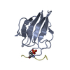 5w7yC S: Starting model for refinement C: citing same article ( |
|---|---|
| Similar structure data |


-Links
 PDBj
PDBj


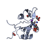
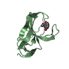
- Assembly
| Deposited unit | 
| ||||||||
|---|---|---|---|---|---|---|---|---|---|
| 1 | 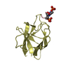
| ||||||||
| 2 | 
| ||||||||
| 3 | 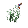
| ||||||||
| 4 | 
| ||||||||
| Unit cell |
|
-Components
| #1: Protein | Mass: 11921.815 Da / Num. of mol.: 4 / Fragment: UNP residues 1-105 Source method: isolated from a genetically manipulated source Source: (gene. exp.) Homo sapiens (human) / Gene: APLF, C2orf13, PALF, XIP1 / Production host:  References: UniProt: Q8IW19, DNA-(apurinic or apyrimidinic site) lyase #2: Protein/peptide | Mass: 1112.878 Da / Num. of mol.: 4 / Fragment: UNP residues 514-522 / Source method: obtained synthetically / Details: bisphosphopeptide of XRCC1 / Source: (synth.) Homo sapiens (human) / References: UniProt: P18887#3: Water | ChemComp-HOH / |  Mass: 18.015 Da / Num. of mol.: 259 / Source method: isolated from a natural source / Formula: H2O Mass: 18.015 Da / Num. of mol.: 259 / Source method: isolated from a natural source / Formula: H2OHas protein modification | Y | |
|---|
-Experimental details
-Experiment
| Experiment | Method: X-RAY DIFFRACTION / Number of used crystals: 1 |
|---|
- Sample preparation
| Crystal | Density Matthews: 2.05 Å3/Da / Density % sol: 40.01 % |
|---|---|
| Crystal grow | Temperature: 293 K / Method: vapor diffusion, sitting drop / pH: 8.5 Details: 0.6mM XRCC1 bisphosphopeptide 0.6mM APLF 0.5M lithium chloride 0.1M Tris 28% PEG 6000 |
-Data collection
| Diffraction | Mean temperature: 100 K |
|---|---|
| Diffraction source | Source: ROTATING ANODE / Type: RIGAKU MICROMAX-007 HF / Wavelength: 1.514 Å |
| Detector | Type: RIGAKU SATURN 944+ / Detector: CCD / Date: Nov 22, 2015 |
| Radiation | Protocol: SINGLE WAVELENGTH / Monochromatic (M) / Laue (L): M / Scattering type: x-ray |
| Radiation wavelength | Wavelength: 1.514 Å / Relative weight: 1 |
| Reflection | Resolution: 2.005→25.497 Å / Num. obs: 28032 / % possible obs: 97.9 % / Redundancy: 4.8 % / Rpim(I) all: 0.031 / Rsym value: 0.065 / Net I/σ(I): 13.4 |
| Reflection shell | Resolution: 2.005→2.05 Å / Redundancy: 2.4 % / Num. unique obs: 1098 / Rpim(I) all: 0.214 / Rsym value: 0.295 / % possible all: 75.3 |
- Processing
| Software |
| |||||||||||||||||||||||||||||||||||||||||||||||||||||||||||||||||||||||||||||
|---|---|---|---|---|---|---|---|---|---|---|---|---|---|---|---|---|---|---|---|---|---|---|---|---|---|---|---|---|---|---|---|---|---|---|---|---|---|---|---|---|---|---|---|---|---|---|---|---|---|---|---|---|---|---|---|---|---|---|---|---|---|---|---|---|---|---|---|---|---|---|---|---|---|---|---|---|---|---|
| Refinement | Method to determine structure: MOLECULAR REPLACEMENT Starting model: 5W7W Resolution: 2.005→25.497 Å / SU ML: 0.23 / Cross valid method: THROUGHOUT / σ(F): 1.34 / Phase error: 26.24
| |||||||||||||||||||||||||||||||||||||||||||||||||||||||||||||||||||||||||||||
| Solvent computation | Shrinkage radii: 0.9 Å / VDW probe radii: 1.11 Å | |||||||||||||||||||||||||||||||||||||||||||||||||||||||||||||||||||||||||||||
| Refinement step | Cycle: LAST / Resolution: 2.005→25.497 Å
| |||||||||||||||||||||||||||||||||||||||||||||||||||||||||||||||||||||||||||||
| Refine LS restraints |
| |||||||||||||||||||||||||||||||||||||||||||||||||||||||||||||||||||||||||||||
| LS refinement shell |
|
