 Movie
Movie Controller

[English] 日本語
 Yorodumi
- PDB-5w2b: Crystal structure of C-terminal domain of Ebola (Reston) nucleopr... -
+ Open data

- Basic information
| Entry | Database: PDB / ID: 5w2b | ||||||
|---|---|---|---|---|---|---|---|
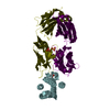
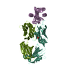
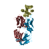
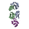
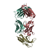
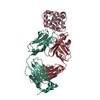
| Title | Crystal structure of C-terminal domain of Ebola (Reston) nucleoprotein in complex with Fab fragment | ||||||
 Components Components |
| ||||||
 Keywords Keywords | viral protein/immune system / Ebola virus / nucleoprotein / Fab / antibody-antigen interaction / VIRAL PROTEIN / viral protein-immune system complex | ||||||
| Function / homology |  Function and homology information Function and homology informationviral RNA genome packaging / helical viral capsid / viral nucleocapsid / host cell cytoplasm / ribonucleoprotein complex / RNA binding Similarity search - Function | ||||||
| Biological species |  Homo sapiens (human) Homo sapiens (human) Reston ebolavirus Reston ebolavirus | ||||||
| Method |  X-RAY DIFFRACTION / SYNCHROTRON / MOLECULAR REPLACEMENT / Resolution: 2.25 Å X-RAY DIFFRACTION / SYNCHROTRON / MOLECULAR REPLACEMENT / Resolution: 2.25 Å | ||||||
 Authors Authors | Radwanska, M.J. / Derewenda, U. / Kossiakoff, A.A. / Derewenda, Z.S. | ||||||
 Citation Citation | Journal: Acta Crystallogr D Struct Biol / Year: 2018 Title: The structure of the C-terminal domain of the nucleoprotein from the Bundibugyo strain of the Ebola virus in complex with a pan-specific synthetic Fab. Authors: Radwanska, M.J. / Jaskolowski, M. / Davydova, E. / Derewenda, U. / Miyake, T. / Engel, D.A. / Kossiakoff, A.A. / Derewenda, Z.S. | ||||||
| History |
|
- Structure visualization
| Structure viewer | Molecule: MolmilJmol/JSmol |
|---|
- Downloads & links
-Download
| PDBx/mmCIF format | 5w2b.cif.gz | 120.6 KB | Display | PDBx/mmCIF format |
|---|---|---|---|---|
| PDB format | pdb5w2b.ent.gz | 91.2 KB | Display | PDB format |
| PDBx/mmJSON format | 5w2b.json.gz | Tree view | PDBx/mmJSON format | |
| Others |  Other downloads Other downloads |
-Validation report
| Summary document | 5w2b_validation.pdf.gz | 439.8 KB | Display | wwPDB validaton report |
|---|---|---|---|---|
| Full document | 5w2b_full_validation.pdf.gz | 443.4 KB | Display | |
| Data in XML | 5w2b_validation.xml.gz | 20.8 KB | Display | |
| Data in CIF | 5w2b_validation.cif.gz | 29 KB | Display | |
| Arichive directory | https://data.pdbj.org/pub/pdb/validation_reports/w2/5w2bftp://data.pdbj.org/pub/pdb/validation_reports/w2/5w2b | HTTPS FTP |
-Related structure data
| Related structure data | 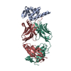 5vkdSC S: Starting model for refinement C: citing same article ( |
|---|---|
| Similar structure data |


-Links
 PDBj
PDBj


- Assembly
| Deposited unit | 
| ||||||||
|---|---|---|---|---|---|---|---|---|---|
| 1 |
| ||||||||
| Unit cell |
|
-Components
| #1: Antibody | Mass: 25216.043 Da / Num. of mol.: 1 Source method: isolated from a genetically manipulated source Source: (gene. exp.) Homo sapiens (human) / Production host:  |
|---|---|
| #2: Antibody | Mass: 23258.783 Da / Num. of mol.: 1 Source method: isolated from a genetically manipulated source Source: (gene. exp.) Homo sapiens (human) / Production host: |
| #3: Protein | Mass: 12462.984 Da / Num. of mol.: 1 Source method: isolated from a genetically manipulated source Source: (gene. exp.) Reston ebolavirus / Strain: Reston-89 / Gene: NP / Production host: |
| #4: Water | ChemComp-HOH /  Mass: 18.015 Da / Num. of mol.: 111 / Source method: isolated from a natural source / Formula: H2O Mass: 18.015 Da / Num. of mol.: 111 / Source method: isolated from a natural source / Formula: H2O |
| Has protein modification | Y |
-Experimental details
-Experiment
| Experiment | Method: X-RAY DIFFRACTION / Number of used crystals: 1 |
|---|
- Sample preparation
| Crystal | Density Matthews: 2.4 Å3/Da / Density % sol: 48.8 % |
|---|---|
| Crystal grow | Temperature: 288 K / Method: vapor diffusion, sitting drop / pH: 7.5 Details: 20 mM xylitol, 20 mM myo-Inositol, 20 mM D-Fructose, 20 mM L-Rhamnose monohydrate, 20 mM D-Sorbitol, 0.1 M BES pH 7.5, 0.1 M triethanolamine (TEA) pH 7.5, 10% w/v PEG 8000, 20% w/v 1,5-Pentanediol |
-Data collection
| Diffraction | Mean temperature: 100 K |
|---|---|
| Diffraction source | Source: SYNCHROTRON / Site: APS  / Beamline: 19-BM / Wavelength: 1 Å / Beamline: 19-BM / Wavelength: 1 Å |
| Detector | Type: ADSC QUANTUM 210r / Detector: CCD / Date: Nov 14, 2015 |
| Radiation | Monochromator: Si(111) / Protocol: SINGLE WAVELENGTH / Monochromatic (M) / Laue (L): M / Scattering type: x-ray |
| Radiation wavelength | Wavelength: 1 Å / Relative weight: 1 |
| Reflection | Resolution: 2.25→50 Å / Num. obs: 28393 / % possible obs: 99.8 % / Redundancy: 7.3 % / Biso Wilson estimate: 50.8 Å2 / Rpim(I) all: 0.034 / Χ2: 1.173 / Net I/σ(I): 3.21 |
| Reflection shell | Resolution: 2.25→2.3 Å / Redundancy: 5 % / Num. unique obs: 1835 / CC1/2: 0.836 / Rpim(I) all: 0.249 |
- Processing
| Software |
| |||||||||||||||||||||||||||||||||||||||||||||||||||||||||||||||||||||||||||||
|---|---|---|---|---|---|---|---|---|---|---|---|---|---|---|---|---|---|---|---|---|---|---|---|---|---|---|---|---|---|---|---|---|---|---|---|---|---|---|---|---|---|---|---|---|---|---|---|---|---|---|---|---|---|---|---|---|---|---|---|---|---|---|---|---|---|---|---|---|---|---|---|---|---|---|---|---|---|---|
| Refinement | Method to determine structure: MOLECULAR REPLACEMENT Starting model: 5VKD Resolution: 2.25→40.623 Å / SU ML: 0.28 / Cross valid method: FREE R-VALUE / σ(F): 1.36 / Phase error: 29.45
| |||||||||||||||||||||||||||||||||||||||||||||||||||||||||||||||||||||||||||||
| Solvent computation | Shrinkage radii: 0.9 Å / VDW probe radii: 1.11 Å | |||||||||||||||||||||||||||||||||||||||||||||||||||||||||||||||||||||||||||||
| Refinement step | Cycle: LAST / Resolution: 2.25→40.623 Å
| |||||||||||||||||||||||||||||||||||||||||||||||||||||||||||||||||||||||||||||
| Refine LS restraints |
| |||||||||||||||||||||||||||||||||||||||||||||||||||||||||||||||||||||||||||||
| LS refinement shell |
|
