 Movie
Movie Controller

[English] 日本語
 Yorodumi
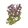
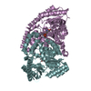
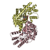
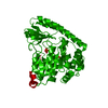
- PDB-5veh: Re-refinement OF THE PDB STRUCTURE 1yiz of Aedes aegypti kynureni... -
+ Open data

- Basic information
| Entry | Database: PDB / ID: 5veh | ||||||
|---|---|---|---|---|---|---|---|
| Title | Re-refinement OF THE PDB STRUCTURE 1yiz of Aedes aegypti kynurenine aminotransferase | ||||||
 Components Components | Kynurenine aminotransferase | ||||||
 Keywords Keywords | TRANSFERASE / Re-refinement / kynurenine aminotransferase / mosquito | ||||||
| Function / homology |  Function and homology information Function and homology information: / kynurenine-glyoxylate transaminase / L-kynurenine:glyoxylate transaminase activity / kynurenine-oxoglutarate transaminase / L-kynurenine:2-oxoglutarate transaminase activity / Transferases; Transferring nitrogenous groups; Transaminases / pyridoxal phosphate binding / mitochondrion Similarity search - Function | ||||||
| Biological species |  | ||||||
| Method |  X-RAY DIFFRACTION / SYNCHROTRON / MOLECULAR REPLACEMENT / Resolution: 1.55 Å X-RAY DIFFRACTION / SYNCHROTRON / MOLECULAR REPLACEMENT / Resolution: 1.55 Å | ||||||
 Authors Authors | Wlodawer, A. / Dauter, Z. / Minor, W. / Stanfield, R. / Porebski, P. / Jaskolski, M. / Pozharski, E. / Weichenberger, C.X. / Rupp, B. | ||||||
 Citation Citation | Journal: FEBS J. / Year: 2005 Title: Crystal structures of Aedes aegypti kynurenine aminotransferase. Authors: Han, Q. / Gao, Y.G. / Robinson, H. / Ding, H. / Wilson, S. / Li, J. | ||||||
| History |
| ||||||
| Remark 0 | This entry reflects an alternative modeling of the original data in 1yiz, determined byHan, Q., ...This entry reflects an alternative modeling of the original data in 1yiz, determined byHan, Q., Gao, Y.G., Robinson, H., Ding, H., Wilson, S., Li, J. | ||||||
| Remark 200 | SEE THE ORIGINAL DATA, ENTRY 1yiz | ||||||
| Remark 280 | SEE THE ORIGINAL DATA, ENTRY 1yiz |
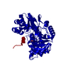
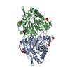
- Structure visualization
| Structure viewer | Molecule: MolmilJmol/JSmol |
|---|
- Downloads & links
-Download
| PDBx/mmCIF format | 5veh.cif.gz | 187.8 KB | Display | PDBx/mmCIF format |
|---|---|---|---|---|
| PDB format | pdb5veh.ent.gz | 151.2 KB | Display | PDB format |
| PDBx/mmJSON format | 5veh.json.gz | Tree view | PDBx/mmJSON format | |
| Others |  Other downloads Other downloads |
-Validation report
| Arichive directory | https://data.pdbj.org/pub/pdb/validation_reports/ve/5vehftp://data.pdbj.org/pub/pdb/validation_reports/ve/5veh | HTTPS FTP |
|---|
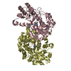
-Related structure data
| Related structure data |  5vepC  5veqC  5verC  5vetC  5vf2C  5vf5C  5vgaC C: citing same article ( |
|---|---|
| Similar structure data |


-Links
 PDBj
PDBj

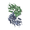
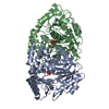
- Assembly
| Deposited unit | 
| ||||||||
|---|---|---|---|---|---|---|---|---|---|
| 1 |
| ||||||||
| Unit cell |
|
-Components
| #1: Protein | Mass: 48390.094 Da / Num. of mol.: 2 Source method: isolated from a genetically manipulated source Source: (gene. exp.)  Spodoptera frugiperda (fall armyworm) / References: UniProt: Q95VY4, UniProt: Q17CS8*PLUS Spodoptera frugiperda (fall armyworm) / References: UniProt: Q95VY4, UniProt: Q17CS8*PLUS#2: Chemical | ChemComp-BR /   Mass: 79.904 Da / Num. of mol.: 12 / Source method: obtained synthetically / Formula: Br Mass: 79.904 Da / Num. of mol.: 12 / Source method: obtained synthetically / Formula: Br#3: Chemical | ChemComp-GOL / |   Mass: 92.094 Da / Num. of mol.: 1 / Source method: obtained synthetically / Formula: C3H8O3 Mass: 92.094 Da / Num. of mol.: 1 / Source method: obtained synthetically / Formula: C3H8O3#4: Water | ChemComp-HOH / |  Mass: 18.015 Da / Num. of mol.: 801 / Source method: isolated from a natural source / Formula: H2O Mass: 18.015 Da / Num. of mol.: 801 / Source method: isolated from a natural source / Formula: H2O |
|---|
-Experimental details
-Experiment
| Experiment | Method: X-RAY DIFFRACTION / Number of used crystals: 1 |
|---|
- Sample preparation
| Crystal | Density Matthews: 2.27 Å3/Da / Density % sol: 45.9 % |
|---|---|
| Crystal grow | Temperature: 277 K / Method: vapor diffusion, hanging drop / Details: PEG 1000, Tris.HCl pH 8.5 |
-Data collection
| Diffraction | Mean temperature: 123 K |
|---|---|
| Diffraction source | Source: SYNCHROTRON / Site: NSLS  / Beamline: X12C / Wavelength: 1.1 Å / Beamline: X12C / Wavelength: 1.1 Å |
| Detector | Type: MARMOSAIC 325 mm CCD / Detector: CCD / Date: Aug 7, 2004 |
| Radiation | Protocol: SINGLE WAVELENGTH / Monochromatic (M) / Laue (L): M / Scattering type: x-ray |
| Radiation wavelength | Wavelength: 1.1 Å / Relative weight: 1 |
| Reflection | Resolution: 1.55→39.3 Å / Num. obs: 128458 / % possible obs: 99.3 % / Redundancy: 7.2 % / Rmerge(I) obs: 0.057 / Net I/σ(I): 2 |
- Processing
| Software |
| |||||||||||||||||||||||||||||||||||||||||||||
|---|---|---|---|---|---|---|---|---|---|---|---|---|---|---|---|---|---|---|---|---|---|---|---|---|---|---|---|---|---|---|---|---|---|---|---|---|---|---|---|---|---|---|---|---|---|---|
| Refinement | Method to determine structure: MOLECULAR REPLACEMENT / Resolution: 1.55→39.3 Å / Cor.coef. Fo:Fc: 0.956 / Cor.coef. Fo:Fc free: 0.941 / Cross valid method: THROUGHOUT / σ(F): 0 / ESU R: 0.082 / ESU R Free: 0.083 Details: HYDROGENS HAVE BEEN USED IF PRESENT IN THE INPUT U VALUES : REFINED INDIVIDUALLY
| |||||||||||||||||||||||||||||||||||||||||||||
| Solvent computation | Ion probe radii: 0.8 Å / Shrinkage radii: 0.8 Å / VDW probe radii: 1.2 Å | |||||||||||||||||||||||||||||||||||||||||||||
| Displacement parameters | Biso max: 51.73 Å2 / Biso mean: 20.548 Å2 / Biso min: 9.56 Å2
| |||||||||||||||||||||||||||||||||||||||||||||
| Refinement step | Cycle: final / Resolution: 1.55→39.3 Å
| |||||||||||||||||||||||||||||||||||||||||||||
| Refine LS restraints |
| |||||||||||||||||||||||||||||||||||||||||||||
| LS refinement shell | Resolution: 1.546→1.586 Å / Rfactor Rfree error: 0 / Total num. of bins used: 20
|
