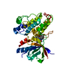
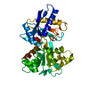
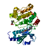
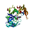
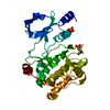
Entry Database : PDB / ID : 5veeTitle PAK4 kinase domain in complex with FRAX486 Serine/threonine-protein kinase PAK 4 Keywords / Function / homology Function Domain/homology Component
/ / / / / / / / / / / / / / / / / / / / / / / / / / / / / / / / / / / / / / / / / / / / / / / / / / / / / / / / / / / / / / / / / / / / / / / / / Biological species Homo sapiens (human)Method / / / Resolution : 2.5 Å Authors Zhang, E.Y. / Ha, B.H. / Boggon, T.J. Funding support Organization Grant number Country National Institutes of Health/National Institute of General Medical Sciences (NIH/NIGMS) R01GM102262 National Institutes of Health/Office of the Director S10OD018007
Journal : Biochim. Biophys. Acta / Year : 2018Title : PAK4 crystal structures suggest unusual kinase conformational movements.Authors : Zhang, E.Y. / Ha, B.H. / Boggon, T.J. History Deposition Apr 4, 2017 Deposition site / Processing site Revision 1.0 Oct 18, 2017 Provider / Type Revision 1.1 Jan 3, 2018 Group / Category Item _citation.journal_volume / _citation.page_first ... _citation.journal_volume / _citation.page_first / _citation.page_last / _citation.year Revision 1.2 Jan 1, 2020 Group / Category / Item Revision 1.3 Feb 26, 2020 Group / Category / reflns_shellItem / _reflns_shell.pdbx_Rpim_I_allRevision 1.4 Oct 4, 2023 Group / Database references / Refinement descriptionCategory chem_comp_atom / chem_comp_bond ... chem_comp_atom / chem_comp_bond / database_2 / pdbx_initial_refinement_model Item / _database_2.pdbx_database_accessionRevision 1.5 Oct 9, 2024 Group / Category / pdbx_modification_feature
Show all Show less
 Movie
Movie Controller


 Open data
 Basic information
 Components
 Keywords
 Function and homology information
 Homo sapiens (human)
 X-RAY DIFFRACTION /
  Authors
 United States, 2items
  Citation
 Structure visualization
 Downloads & links
 Other downloads
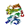


 PDBj
PDBj


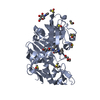
 Assembly
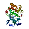


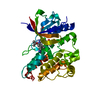
 Mass: 513.394 Da / Num. of mol.: 1 / Source method: obtained synthetically / Formula: C25H23Cl2FN6O
 Sample preparation
 Processing