 Movie
Movie Controller

[English] 日本語
 Yorodumi
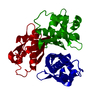
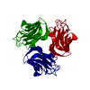
- PDB-5vbx: Crystal structure of holo-[acyl-carrier-protein] synthase (AcpS) ... -
+ Open data

- Basic information
| Entry | Database: PDB / ID: 5vbx | |||||||||
|---|---|---|---|---|---|---|---|---|---|---|
| Title | Crystal structure of holo-[acyl-carrier-protein] synthase (AcpS) from Escherichia coli | |||||||||
 Components Components | Holo-[acyl-carrier-protein] synthase | |||||||||
 Keywords Keywords | TRANSFERASE / Trimer | |||||||||
| Function / homology |  Function and homology information Function and homology informationholo-[acyl-carrier-protein] synthase / holo-[acyl-carrier-protein] synthase activity / fatty acid biosynthetic process / transferase activity / magnesium ion binding / cytoplasm Similarity search - Function | |||||||||
| Biological species |  | |||||||||
| Method |  X-RAY DIFFRACTION / SYNCHROTRON / MOLECULAR REPLACEMENT / Resolution: 2.05 Å X-RAY DIFFRACTION / SYNCHROTRON / MOLECULAR REPLACEMENT / Resolution: 2.05 Å | |||||||||
 Authors Authors | Marcella, A.M. / Barb, A.W. | |||||||||
| Funding support |  United States, 2items United States, 2items
| |||||||||
 Citation Citation | Journal: J. Mol. Biol. / Year: 2017 Title: Structure, High Affinity, and Negative Cooperativity of the Escherichia coli Holo-(Acyl Carrier Protein):Holo-(Acyl Carrier Protein) Synthase Complex. Authors: Marcella, A.M. / Culbertson, S.J. / Shogren-Knaak, M.A. / Barb, A.W. | |||||||||
| History |
|
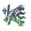
- Structure visualization
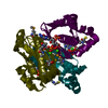
| Structure viewer | Molecule: MolmilJmol/JSmol |
|---|
- Downloads & links
-Download
| PDBx/mmCIF format | 5vbx.cif.gz | 91.3 KB | Display | PDBx/mmCIF format |
|---|---|---|---|---|
| PDB format | pdb5vbx.ent.gz | 69.2 KB | Display | PDB format |
| PDBx/mmJSON format | 5vbx.json.gz | Tree view | PDBx/mmJSON format | |
| Others |  Other downloads Other downloads |
-Validation report
| Arichive directory | https://data.pdbj.org/pub/pdb/validation_reports/vb/5vbxftp://data.pdbj.org/pub/pdb/validation_reports/vb/5vbx | HTTPS FTP |
|---|
-Related structure data
| Related structure data |  5vcbC  1f7tS S: Starting model for refinement C: citing same article ( |
|---|---|
| Similar structure data |


-Links
 PDBj
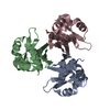
PDBj- Assembly
| Deposited unit | 
| ||||||||
|---|---|---|---|---|---|---|---|---|---|
| 1 |
| ||||||||
| Unit cell |
| ||||||||
| Components on special symmetry positions |
|
-Components
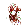
| #1: Protein | Mass: 14074.297 Da / Num. of mol.: 3 Source method: isolated from a genetically manipulated source Source: (gene. exp.) Strain: K12 / Gene: acpS, dpj, b2563, JW2547 / Production host: References: UniProt: P24224, holo-[acyl-carrier-protein] synthase #2: Chemical | ChemComp-EDO /   Mass: 62.068 Da / Num. of mol.: 8 / Source method: obtained synthetically / Formula: C2H6O2 Mass: 62.068 Da / Num. of mol.: 8 / Source method: obtained synthetically / Formula: C2H6O2#3: Chemical | ChemComp-NA /   Mass: 22.990 Da / Num. of mol.: 9 / Source method: obtained synthetically / Formula: Na Mass: 22.990 Da / Num. of mol.: 9 / Source method: obtained synthetically / Formula: Na#4: Water | ChemComp-HOH / |  Mass: 18.015 Da / Num. of mol.: 156 / Source method: isolated from a natural source / Formula: H2O Mass: 18.015 Da / Num. of mol.: 156 / Source method: isolated from a natural source / Formula: H2O |
|---|
-Experimental details
-Experiment
| Experiment | Method: X-RAY DIFFRACTION / Number of used crystals: 1 |
|---|
- Sample preparation
| Crystal | Density Matthews: 2.33 Å3/Da / Density % sol: 47.26 % |
|---|---|
| Crystal grow | Temperature: 293 K / Method: vapor diffusion, hanging drop / pH: 7 Details: 1.8-2.2 M Sodium Formate 1.8-2.2 M Sodium Nitrate 100 mM HEPES pH 7.0 |
-Data collection
| Diffraction | Mean temperature: 100 K |
|---|---|
| Diffraction source | Source: SYNCHROTRON / Site: APS / Beamline: 23-ID-B / Wavelength: 1.003 Å |
| Detector | Type: MARMOSAIC 300 mm CCD / Detector: CCD / Date: Feb 21, 2016 |
| Radiation | Protocol: SINGLE WAVELENGTH / Monochromatic (M) / Laue (L): M / Scattering type: x-ray |
| Radiation wavelength | Wavelength: 1.003 Å / Relative weight: 1 |
| Reflection | Resolution: 2.05→29.598 Å / Num. obs: 24465 / % possible obs: 100 % / Redundancy: 4.2 % / CC1/2: 0.996 / Rmerge(I) obs: 0.1458 / Net I/σ(I): 10.24 |
| Reflection shell | Highest resolution: 2.05 Å / Redundancy: 3.9 % / Num. unique obs: 2704 / CC1/2: 0.538 / % possible all: 99.9 |
- Processing
| Software |
| |||||||||||||||||||||||||||||||||||||||||||||||||||||||||||||||
|---|---|---|---|---|---|---|---|---|---|---|---|---|---|---|---|---|---|---|---|---|---|---|---|---|---|---|---|---|---|---|---|---|---|---|---|---|---|---|---|---|---|---|---|---|---|---|---|---|---|---|---|---|---|---|---|---|---|---|---|---|---|---|---|---|
| Refinement | Method to determine structure: MOLECULAR REPLACEMENT Starting model: 1F7T Resolution: 2.05→29.598 Å / SU ML: 0.28 / Cross valid method: THROUGHOUT / σ(F): 1.37 / Phase error: 25.83 / Stereochemistry target values: ML
| |||||||||||||||||||||||||||||||||||||||||||||||||||||||||||||||
| Solvent computation | Shrinkage radii: 0.9 Å / VDW probe radii: 1.11 Å / Solvent model: FLAT BULK SOLVENT MODEL | |||||||||||||||||||||||||||||||||||||||||||||||||||||||||||||||
| Refinement step | Cycle: LAST / Resolution: 2.05→29.598 Å
| |||||||||||||||||||||||||||||||||||||||||||||||||||||||||||||||
| Refine LS restraints |
| |||||||||||||||||||||||||||||||||||||||||||||||||||||||||||||||
| LS refinement shell |
| |||||||||||||||||||||||||||||||||||||||||||||||||||||||||||||||
| Refinement TLS params. | Method: refined / Origin x: 127.6352 Å / Origin y: -9.4766 Å / Origin z: 245.3615 Å
| |||||||||||||||||||||||||||||||||||||||||||||||||||||||||||||||
| Refinement TLS group | Selection details: all |
