 Movie
Movie Controller

[English] 日本語
 Yorodumi
- PDB-5v7s: Crystal structure of Influenza A virus matrix protein M1 (NLS-88E... -
+ Open data

- Basic information
| Entry | Database: PDB / ID: 5v7s | |||||||||
|---|---|---|---|---|---|---|---|---|---|---|
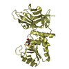
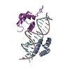
| Title | Crystal structure of Influenza A virus matrix protein M1 (NLS-88E, pH 6.2) | |||||||||
 Components Components | Matrix protein 1 | |||||||||
 Keywords Keywords | VIRAL PROTEIN / Influenza A / Matrix protein / NLS-88E mutant / pH 6.2 | |||||||||
| Function / homology |  Function and homology information Function and homology informationviral budding from plasma membrane / structural constituent of virion / host cell nucleus / virion membrane / RNA binding Similarity search - Function | |||||||||
| Biological species |   Influenza A virus Influenza A virus | |||||||||
| Method |  X-RAY DIFFRACTION / MOLECULAR REPLACEMENT / Resolution: 2.5 Å X-RAY DIFFRACTION / MOLECULAR REPLACEMENT / Resolution: 2.5 Å | |||||||||
 Authors Authors | Musayev, F.N. / Safo, M.K. / Althufairi, B. / Desai, U.R. / Xie, H. / Mosier, P.D. / Chiang, M.-J. / Zhou, Q. | |||||||||
| Funding support |  United States, 2items United States, 2items
| |||||||||
 Citation Citation | Journal: Emerg Microbes Infect / Year: 2017 Title: Maintaining pH-dependent conformational flexibility of M1 is critical for efficient influenza A virus replication. Authors: Chiang, M.J. / Musayev, F.N. / Kosikova, M. / Lin, Z. / Gao, Y. / Mosier, P.D. / Althufairi, B. / Ye, Z. / Zhou, Q. / Desai, U.R. / Xie, H. / Safo, M.K. | |||||||||
| History |
|
- Structure visualization
| Structure viewer | Molecule: MolmilJmol/JSmol |
|---|
- Downloads & links
-Download
| PDBx/mmCIF format | 5v7s.cif.gz | 102.6 KB | Display | PDBx/mmCIF format |
|---|---|---|---|---|
| PDB format | pdb5v7s.ent.gz | 77.9 KB | Display | PDB format |
| PDBx/mmJSON format | 5v7s.json.gz | Tree view | PDBx/mmJSON format | |
| Others |  Other downloads Other downloads |
-Validation report
| Arichive directory | https://data.pdbj.org/pub/pdb/validation_reports/v7/5v7sftp://data.pdbj.org/pub/pdb/validation_reports/v7/5v7s | HTTPS FTP |
|---|
-Related structure data
| Related structure data |  5v6gC  5v7bC  5v8aC  1ea3S C: citing same article ( S: Starting model for refinement |
|---|---|
| Similar structure data |

-Links
 PDBj
PDBj

- Assembly
| Deposited unit | 
| ||||||||
|---|---|---|---|---|---|---|---|---|---|
| 1 | 
| ||||||||
| 2 | 
| ||||||||
| Unit cell |
|
-Components
| #1: Protein | Mass: 19069.975 Da / Num. of mol.: 3 / Mutation: G88E, R101S, R105S Source method: isolated from a genetically manipulated source Source: (gene. exp.) Influenza A virus (strain A/Wilson-Smith/1933 H1N1)Strain: A/Wilson-Smith/1933 H1N1 / Plasmid: pET30a / Production host:  #2: Chemical |   Mass: 94.971 Da / Num. of mol.: 2 / Source method: obtained synthetically / Formula: PO4 Mass: 94.971 Da / Num. of mol.: 2 / Source method: obtained synthetically / Formula: PO4#3: Water | ChemComp-HOH / |  Mass: 18.015 Da / Num. of mol.: 13 / Source method: isolated from a natural source / Formula: H2O Mass: 18.015 Da / Num. of mol.: 13 / Source method: isolated from a natural source / Formula: H2O |
|---|
-Experimental details
-Experiment
| Experiment | Method: X-RAY DIFFRACTION / Number of used crystals: 1 |
|---|
- Sample preparation
| Crystal | Density Matthews: 2.04 Å3/Da / Density % sol: 39.8 % |
|---|---|
| Crystal grow | Temperature: 293 K / Method: vapor diffusion, sitting drop / pH: 6.2 Details: Protein solution: 8 mg/ml in 50mM K2HPO4/KH2PO4/H3PO4, 0.2M NaCl, 10mM bME, pH 3.2 Reservoir solution: 0.1M Tris-HCl, pH 8.2, 8% PEG-8K |
-Data collection
| Diffraction | Mean temperature: 100 K |
|---|---|
| Diffraction source | Source: ROTATING ANODE / Type: RIGAKU MICROMAX-007 / Wavelength: 1.5418 Å |
| Detector | Type: RIGAKU RAXIS IV++ / Detector: IMAGE PLATE / Date: Aug 20, 2015 / Details: Rigaku varimax confocal |
| Radiation | Protocol: SINGLE WAVELENGTH / Monochromatic (M) / Laue (L): M / Scattering type: x-ray |
| Radiation wavelength | Wavelength: 1.5418 Å / Relative weight: 1 |
| Reflection | Resolution: 2.5→26.23 Å / Num. obs: 14742 / % possible obs: 90.7 % / Redundancy: 5.37 % / Biso Wilson estimate: 55.06 Å2 / Rmerge(I) obs: 0.077 / Net I/σ(I): 12.1 |
| Reflection shell | Resolution: 2.5→2.59 Å / Redundancy: 5.51 % / Rmerge(I) obs: 0.449 / Mean I/σ(I) obs: 3.6 / Num. unique obs: 1473 / % possible all: 91.9 |
- Processing
| Software |
| |||||||||||||||||||||||||||||||||||
|---|---|---|---|---|---|---|---|---|---|---|---|---|---|---|---|---|---|---|---|---|---|---|---|---|---|---|---|---|---|---|---|---|---|---|---|---|
| Refinement | Method to determine structure: MOLECULAR REPLACEMENT Starting model: 1EA3 Resolution: 2.5→26.23 Å / SU ML: 0.49 / Cross valid method: THROUGHOUT / σ(F): 1.34 / Phase error: 35.93
| |||||||||||||||||||||||||||||||||||
| Solvent computation | Shrinkage radii: 0.9 Å / VDW probe radii: 1.11 Å | |||||||||||||||||||||||||||||||||||
| Refinement step | Cycle: LAST / Resolution: 2.5→26.23 Å
| |||||||||||||||||||||||||||||||||||
| Refine LS restraints |
| |||||||||||||||||||||||||||||||||||
| LS refinement shell |
|
