 Movie
Movie Controller

[English] 日本語
 Yorodumi
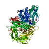
- PDB-5v12: Crystal structure of Carbon Sulfoxide lyase, Egt2 Y134F with sulf... -
+ Open data

- Basic information
| Entry | Database: PDB / ID: 5v12 | ||||||||||||
|---|---|---|---|---|---|---|---|---|---|---|---|---|---|
| Title | Crystal structure of Carbon Sulfoxide lyase, Egt2 Y134F with sulfenic acid intermediate | ||||||||||||
 Components Components | (Hercynylcysteine sulfoxide lyase) x 2 | ||||||||||||
 Keywords Keywords | LYASE / C-S lyase / PLP dependent | ||||||||||||
| Function / homology |  Function and homology information Function and homology informationLyases; Carbon-sulfur lyases / hercynylcysteine sulfoxide lyase activity (ergothioneine-forming) / nucleus / cytoplasm Similarity search - Function | ||||||||||||
| Biological species |  Neurospora crassa OR74A (fungus) Neurospora crassa OR74A (fungus) | ||||||||||||
| Method |  X-RAY DIFFRACTION / SYNCHROTRON / MOLECULAR REPLACEMENT / Resolution: 2.451 Å X-RAY DIFFRACTION / SYNCHROTRON / MOLECULAR REPLACEMENT / Resolution: 2.451 Å | ||||||||||||
 Authors Authors | Irani, S. / Zhang, Y. | ||||||||||||
 Citation Citation | Journal: Cell Chem Biol / Year: 2018 Title: Snapshots of C-S Cleavage in Egt2 Reveals Substrate Specificity and Reaction Mechanism. Authors: Irani, S. / Naowarojna, N. / Tang, Y. / Kathuria, K.R. / Wang, S. / Dhembi, A. / Lee, N. / Yan, W. / Lyu, H. / Costello, C.E. / Liu, P. / Zhang, Y.J. | ||||||||||||
| History |
|
- Structure visualization
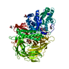
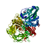
| Structure viewer | Molecule: MolmilJmol/JSmol |
|---|
- Downloads & links
-Download
| PDBx/mmCIF format | 5v12.cif.gz | 696.5 KB | Display | PDBx/mmCIF format |
|---|---|---|---|---|
| PDB format | pdb5v12.ent.gz | 570.6 KB | Display | PDB format |
| PDBx/mmJSON format | 5v12.json.gz | Tree view | PDBx/mmJSON format | |
| Others |  Other downloads Other downloads |
-Validation report
| Summary document | 5v12_validation.pdf.gz | 565.3 KB | Display | wwPDB validaton report |
|---|---|---|---|---|
| Full document | 5v12_full_validation.pdf.gz | 618.5 KB | Display | |
| Data in XML | 5v12_validation.xml.gz | 126 KB | Display | |
| Data in CIF | 5v12_validation.cif.gz | 167.4 KB | Display | |
| Arichive directory | https://data.pdbj.org/pub/pdb/validation_reports/v1/5v12ftp://data.pdbj.org/pub/pdb/validation_reports/v1/5v12 | HTTPS FTP |
-Related structure data
| Related structure data | 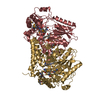 5utsSC 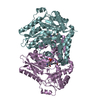 5v1xC S: Starting model for refinement C: citing same article ( |
|---|---|
| Similar structure data |


-Links
 PDBj
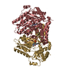
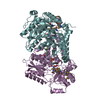
PDBj- Assembly
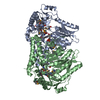
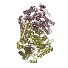
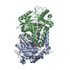
| Deposited unit | 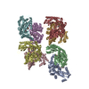
| ||||||||
|---|---|---|---|---|---|---|---|---|---|
| 1 | 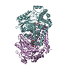
| ||||||||
| 2 | 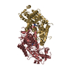
| ||||||||
| 3 | 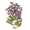
| ||||||||
| 4 | 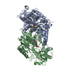
| ||||||||
| Unit cell |
| ||||||||
| Details | Dimer as determined by gel filtration |
-Components
| #1: Protein | Mass: 56426.262 Da / Num. of mol.: 7 Source method: isolated from a genetically manipulated source Source: (gene. exp.) Neurospora crassa OR74A (fungus)Strain: ATCC 24698 / 74-OR23-1A / CBS 708.71 / DSM 1257 / FGSC 987 Gene: egt-2, NCU11365 / Production host:  #2: Protein | | Mass: 56513.340 Da / Num. of mol.: 1 Source method: isolated from a genetically manipulated source Source: (gene. exp.) Neurospora crassa OR74A (fungus)Strain: ATCC 24698 / 74-OR23-1A / CBS 708.71 / DSM 1257 / FGSC 987 Gene: egt-2, NCU11365 / Production host: #3: Chemical | ChemComp-FMT /   Mass: 46.025 Da / Num. of mol.: 8 / Source method: obtained synthetically / Formula: CH2O2 Mass: 46.025 Da / Num. of mol.: 8 / Source method: obtained synthetically / Formula: CH2O2#4: Chemical | 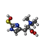  Mass: 246.307 Da / Num. of mol.: 2 / Source method: obtained synthetically / Formula: C9H16N3O3S Mass: 246.307 Da / Num. of mol.: 2 / Source method: obtained synthetically / Formula: C9H16N3O3S#5: Water | ChemComp-HOH / |  Mass: 18.015 Da / Num. of mol.: 412 / Source method: isolated from a natural source / Formula: H2O Mass: 18.015 Da / Num. of mol.: 412 / Source method: isolated from a natural source / Formula: H2O |
|---|
-Experimental details
-Experiment
| Experiment | Method: X-RAY DIFFRACTION / Number of used crystals: 1 |
|---|
- Sample preparation
| Crystal | Density Matthews: 2.82 Å3/Da / Density % sol: 56.33 % |
|---|---|
| Crystal grow | Temperature: 277 K / Method: vapor diffusion, sitting drop Details: 30% PEG 3350, 50mM HEPES pH 7.5, 200mM sodium formate |
-Data collection
| Diffraction | Mean temperature: 100 K |
|---|---|
| Diffraction source | Source: SYNCHROTRON / Site: APS  / Beamline: 23-ID-D / Wavelength: 1.0332 Å / Beamline: 23-ID-D / Wavelength: 1.0332 Å |
| Detector | Type: DECTRIS PILATUS3 6M / Detector: PIXEL / Date: Feb 17, 2016 |
| Radiation | Protocol: SINGLE WAVELENGTH / Monochromatic (M) / Laue (L): M / Scattering type: x-ray |
| Radiation wavelength | Wavelength: 1.0332 Å / Relative weight: 1 |
| Reflection | Resolution: 2.45→50 Å / Num. obs: 146021 / % possible obs: 91.2 % / Redundancy: 2.9 % / Net I/σ(I): 8.46 |
- Processing
| Software |
| |||||||||||||||||||||||||||||||||||||||||||||||||||||||||||||||||||||||||||||||||||||||||||||||||||||||||
|---|---|---|---|---|---|---|---|---|---|---|---|---|---|---|---|---|---|---|---|---|---|---|---|---|---|---|---|---|---|---|---|---|---|---|---|---|---|---|---|---|---|---|---|---|---|---|---|---|---|---|---|---|---|---|---|---|---|---|---|---|---|---|---|---|---|---|---|---|---|---|---|---|---|---|---|---|---|---|---|---|---|---|---|---|---|---|---|---|---|---|---|---|---|---|---|---|---|---|---|---|---|---|---|---|---|---|
| Refinement | Method to determine structure: MOLECULAR REPLACEMENT Starting model: 5UTS Resolution: 2.451→49.577 Å / SU ML: 0.36 / Cross valid method: FREE R-VALUE / σ(F): 1.34 / Phase error: 32.33
| |||||||||||||||||||||||||||||||||||||||||||||||||||||||||||||||||||||||||||||||||||||||||||||||||||||||||
| Solvent computation | Shrinkage radii: 0.9 Å / VDW probe radii: 1.11 Å | |||||||||||||||||||||||||||||||||||||||||||||||||||||||||||||||||||||||||||||||||||||||||||||||||||||||||
| Refinement step | Cycle: LAST / Resolution: 2.451→49.577 Å
| |||||||||||||||||||||||||||||||||||||||||||||||||||||||||||||||||||||||||||||||||||||||||||||||||||||||||
| Refine LS restraints |
| |||||||||||||||||||||||||||||||||||||||||||||||||||||||||||||||||||||||||||||||||||||||||||||||||||||||||
| LS refinement shell |
|
