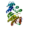
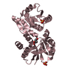
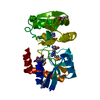
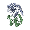
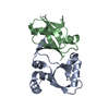
Nucleotidyl transferase AbiEii toxin, Type IV TA system / Nucleotidyl transferase AbiEii toxin, Type IV TA system / Nucleotidyltransferase superfamily / metal ion binding / Uncharacterized protein
Function and homology information
Biological species
Paracoccidioides brasiliensis
Method
X-RAY DIFFRACTION / SYNCHROTRON / SAD / Resolution: 1.86 Å
Mass: 18.015 Da / Num. of mol.: 247 / Source method: isolated from a natural source / Formula: H2O
-
Experimental details
-
Experiment
Experiment
Method: X-RAY DIFFRACTION / Number of used crystals: 2
-
Sample preparation
Crystal
ID
Density Matthews (Å3/Da)
Density % sol (%)
Description
1
2.1
41.5
thin and long rod-like crystals
2
2.1
41.5
thin and long rod-like crystals
Crystal grow
Temperature (K)
Crystal-ID
Method
Details
PH range
293.15
1
vapor diffusion, hanging drop
0.25 M MgCl2, 25% PEG 4000, 0.1 M Tris-HCl pH 7.0 - 8,0
7.0 - 8.0
293.15
2
vapor diffusion, hanging drop
0.25 M MgCl2, 25% PEG 4000, 0.1 M Tris-HCl pH 7.0 - 8,0 Crystal was incubated in a cryo-soaking solution containing 100 mM NaI and 10% (v/v) ethylene glycol
Method to determine structure: SAD / Resolution: 1.86→47.09 Å / Cor.coef. Fo:Fc: 0.959 / Cor.coef. Fo:Fc free: 0.92 / SU B: 3.152 / SU ML: 0.095 / Cross valid method: THROUGHOUT / ESU R: 0.141 / ESU R Free: 0.144 / Details: HYDROGENS HAVE BEEN ADDED IN THE RIDING POSITIONS
Rfactor
Num. reflection
% reflection
Selection details
Rfree
0.22168
895
5 %
RANDOM
Rwork
0.15909
-
-
-
obs
0.16216
16999
97.85 %
-
Solvent computation
Ion probe radii: 0.8 Å / Shrinkage radii: 0.8 Å / VDW probe radii: 1.2 Å
 Movie
Movie Controller

 Yorodumi
 Open data
 Basic information
 Components
 Keywords
 Function and homology information
 X-RAY DIFFRACTION /
  Authors
 Brazil, 1items
  Citation
 Structure visualization
 Downloads & links
 Other downloads
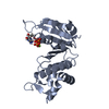


 PDBj
PDBj Assembly
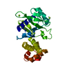
 Paracoccidioides brasiliensis (strain Pb18) (fungus)

 Mass: 24.305 Da / Num. of mol.: 3 / Source method: obtained synthetically / Formula: Mg
 Mass: 18.015 Da / Num. of mol.: 247 / Source method: isolated from a natural source / Formula: H2O
 Sample preparation
 Processing