 Movie
Movie Controller

[English] 日本語
 Yorodumi
- PDB-5utl: Mutant Structures of Streptococcus Agalactiae GBS Glyceraldehyde-... -
+ Open data

- Basic information
| Entry | Database: PDB / ID: 5utl | ||||||
|---|---|---|---|---|---|---|---|
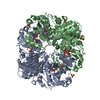
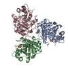
| Title | Mutant Structures of Streptococcus Agalactiae GBS Glyceraldehyde-3-Phosphate Dehydrogenase (GAPDH) | ||||||
 Components Components | Glyceraldehyde-3-phosphate dehydrogenase | ||||||
 Keywords Keywords | OXIDOREDUCTASE / NAD / GAPDH / GLYCOLYSIS | ||||||
| Function / homology |  Function and homology information Function and homology informationOxidoreductases; Acting on the aldehyde or oxo group of donors; With NAD+ or NADP+ as acceptor / glyceraldehyde-3-phosphate dehydrogenase (NAD+) (phosphorylating) activity / glycolytic process / glucose metabolic process / NAD binding / NADP binding / metal ion binding / identical protein binding Similarity search - Function | ||||||
| Biological species |  Streptococcus agalactiae (bacteria) Streptococcus agalactiae (bacteria) | ||||||
| Method |  X-RAY DIFFRACTION / SYNCHROTRON / MOLECULAR REPLACEMENT / Resolution: 1.92 Å X-RAY DIFFRACTION / SYNCHROTRON / MOLECULAR REPLACEMENT / Resolution: 1.92 Å | ||||||
 Authors Authors | Schormann, N. / Ulett, G.C. / Chattopadhyay, D. | ||||||
| Funding support |  Australia, 1items Australia, 1items
| ||||||
 Citation Citation | Journal: To Be Published Title: Mutant Structures of Streptococcus agalactiae GAPDH Authors: Schormann, N. / Ulett, G.C. / Chattopadhyay, D. | ||||||
| History |
|
- Structure visualization
| Structure viewer | Molecule: MolmilJmol/JSmol |
|---|
- Downloads & links
-Download
| PDBx/mmCIF format | 5utl.cif.gz | 272.6 KB | Display | PDBx/mmCIF format |
|---|---|---|---|---|
| PDB format | pdb5utl.ent.gz | 218.5 KB | Display | PDB format |
| PDBx/mmJSON format | 5utl.json.gz | Tree view | PDBx/mmJSON format | |
| Others |  Other downloads Other downloads |
-Validation report
| Arichive directory | https://data.pdbj.org/pub/pdb/validation_reports/ut/5utlftp://data.pdbj.org/pub/pdb/validation_reports/ut/5utl | HTTPS FTP |
|---|
-Related structure data
| Related structure data |  5utmC  5jy6S S: Starting model for refinement C: citing same article ( |
|---|---|
| Similar structure data |

-Links
 PDBj
PDBj
- Assembly
| Deposited unit | 
| ||||||||
|---|---|---|---|---|---|---|---|---|---|
| 1 |
| ||||||||
| Unit cell |
|
-Components
| #1: Protein | Mass: 38199.895 Da / Num. of mol.: 4 / Mutation: Q300L Source method: isolated from a genetically manipulated source Source: (gene. exp.) Streptococcus agalactiae (bacteria)Gene: gapA, gap, AMR84_09125, AX245_09885, BBP08_10800, DX05_09270, EN72_09590, ERS039640_00200, RDF_1710, TH70_1537 Plasmid: PET15b / Production host: References: UniProt: Q9ALW2, Oxidoreductases; Acting on the aldehyde or oxo group of donors; With NAD+ or NADP+ as acceptor #2: Chemical | ChemComp-NAD /   Mass: 663.425 Da / Num. of mol.: 4 / Source method: obtained synthetically / Formula: C21H27N7O14P2 / Comment: NAD*YM Mass: 663.425 Da / Num. of mol.: 4 / Source method: obtained synthetically / Formula: C21H27N7O14P2 / Comment: NAD*YM#3: Chemical | ChemComp-MG /   Mass: 24.305 Da / Num. of mol.: 4 / Source method: obtained synthetically / Formula: Mg Mass: 24.305 Da / Num. of mol.: 4 / Source method: obtained synthetically / Formula: Mg#4: Water | ChemComp-HOH / |  Mass: 18.015 Da / Num. of mol.: 473 / Source method: isolated from a natural source / Formula: H2O Mass: 18.015 Da / Num. of mol.: 473 / Source method: isolated from a natural source / Formula: H2O |
|---|
-Experimental details
-Experiment
| Experiment | Method: X-RAY DIFFRACTION / Number of used crystals: 1 |
|---|
- Sample preparation
| Crystal | Density Matthews: 2.09 Å3/Da / Density % sol: 41.16 % |
|---|---|
| Crystal grow | Temperature: 295 K / Method: vapor diffusion, hanging drop / pH: 6.5 / Details: 20-28% PEG4000, 0.1M Mes |
-Data collection
| Diffraction | Mean temperature: 100 K |
|---|---|
| Diffraction source | Source: SYNCHROTRON / Site: APS  / Beamline: 24-ID-C / Wavelength: 0.9791 Å / Beamline: 24-ID-C / Wavelength: 0.9791 Å |
| Detector | Type: DECTRIS PILATUS 6M / Detector: PIXEL / Date: Nov 12, 2015 / Details: Mirrors |
| Radiation | Monochromator: Cryo cooled double crystal Si / Protocol: SINGLE WAVELENGTH / Monochromatic (M) / Laue (L): M / Scattering type: x-ray |
| Radiation wavelength | Wavelength: 0.9791 Å / Relative weight: 1 |
| Reflection | Resolution: 1.92→108.41 Å / Num. obs: 92332 / % possible obs: 95.8 % / Redundancy: 3.4 % / Biso Wilson estimate: 30 Å2 / CC1/2: 0.992 / Rmerge(I) obs: 0.098 / Rpim(I) all: 0.061 / Rrim(I) all: 0.116 / Net I/σ(I): 11.9 |
| Reflection shell | Resolution: 1.92→1.95 Å / Redundancy: 1.7 % / Rmerge(I) obs: 0.768 / Mean I/σ(I) obs: 1 / Num. unique obs: 3191 / CC1/2: 0.444 / Rpim(I) all: 0.736 / Rrim(I) all: 1.066 / % possible all: 66.6 |
- Processing
| Software |
| ||||||||||||||||||||||||||||||||||||||||||||||||||||||||||||
|---|---|---|---|---|---|---|---|---|---|---|---|---|---|---|---|---|---|---|---|---|---|---|---|---|---|---|---|---|---|---|---|---|---|---|---|---|---|---|---|---|---|---|---|---|---|---|---|---|---|---|---|---|---|---|---|---|---|---|---|---|---|
| Refinement | Method to determine structure: MOLECULAR REPLACEMENT Starting model: 5JY6 Resolution: 1.92→86.87 Å / Cor.coef. Fo:Fc: 0.954 / Cor.coef. Fo:Fc free: 0.944 / SU B: 4.442 / SU ML: 0.12 / Cross valid method: THROUGHOUT / σ(F): 0 / ESU R: 0.193 / ESU R Free: 0.152 Details: HYDROGENS HAVE BEEN ADDED IN THE RIDING POSITIONS U VALUES : REFINED INDIVIDUALLY
| ||||||||||||||||||||||||||||||||||||||||||||||||||||||||||||
| Solvent computation | Ion probe radii: 0.8 Å / Shrinkage radii: 0.8 Å / VDW probe radii: 1.2 Å | ||||||||||||||||||||||||||||||||||||||||||||||||||||||||||||
| Displacement parameters | Biso max: 69.67 Å2 / Biso mean: 30.552 Å2 / Biso min: 14.04 Å2
| ||||||||||||||||||||||||||||||||||||||||||||||||||||||||||||
| Refinement step | Cycle: final / Resolution: 1.92→86.87 Å
| ||||||||||||||||||||||||||||||||||||||||||||||||||||||||||||
| Refine LS restraints |
| ||||||||||||||||||||||||||||||||||||||||||||||||||||||||||||
| LS refinement shell | Resolution: 1.92→1.96 Å
|
