 Movie
Movie Controller

[English] 日本語
 Yorodumi
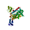
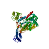
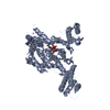
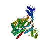
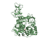
- PDB-5uqt: Clostridium difficile Toxin B (TcdB) glucosyltransferase domain c... -
+ Open data

- Basic information
| Entry | Database: PDB / ID: 5uqt | ||||||||||||
|---|---|---|---|---|---|---|---|---|---|---|---|---|---|
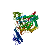
| Title | Clostridium difficile Toxin B (TcdB) glucosyltransferase domain co-crystallized with apigenin | ||||||||||||
 Components Components | Toxin B | ||||||||||||
 Keywords Keywords | TRANSFERASE / glucosyltransferase / toxin | ||||||||||||
| Function / homology |  Function and homology information Function and homology informationsymbiont-mediated perturbation of host actin cytoskeleton via filamentous actin depolymerization / glucosyltransferase activity / Transferases; Glycosyltransferases; Hexosyltransferases / host cell cytosol / cysteine-type peptidase activity / host cell endosome membrane / toxin activity / Hydrolases; Acting on peptide bonds (peptidases); Cysteine endopeptidases / lipid binding / host cell plasma membrane ...symbiont-mediated perturbation of host actin cytoskeleton via filamentous actin depolymerization / glucosyltransferase activity / Transferases; Glycosyltransferases; Hexosyltransferases / host cell cytosol / cysteine-type peptidase activity / host cell endosome membrane / toxin activity / Hydrolases; Acting on peptide bonds (peptidases); Cysteine endopeptidases / lipid binding / host cell plasma membrane / proteolysis / extracellular region / metal ion binding Similarity search - Function | ||||||||||||
| Biological species |  Clostridioides difficile (bacteria) Clostridioides difficile (bacteria) | ||||||||||||
| Method |  X-RAY DIFFRACTION / SYNCHROTRON / MOLECULAR REPLACEMENT / Resolution: 2.75 Å X-RAY DIFFRACTION / SYNCHROTRON / MOLECULAR REPLACEMENT / Resolution: 2.75 Å | ||||||||||||
 Authors Authors | Alvin, J.W. / Lacy, D.B. | ||||||||||||
| Funding support |  United States, 3items United States, 3items
| ||||||||||||
 Citation Citation | Journal: J. Struct. Biol. / Year: 2017 Title: Clostridium difficile toxin glucosyltransferase domains in complex with a non-hydrolyzable UDP-glucose analogue. Authors: Alvin, J.W. / Lacy, D.B. | ||||||||||||
| History |
|
- Structure visualization
| Structure viewer | Molecule: MolmilJmol/JSmol |
|---|
- Downloads & links
-Download
| PDBx/mmCIF format | 5uqt.cif.gz | 454.4 KB | Display | PDBx/mmCIF format |
|---|---|---|---|---|
| PDB format | pdb5uqt.ent.gz | 377.5 KB | Display | PDB format |
| PDBx/mmJSON format | 5uqt.json.gz | Tree view | PDBx/mmJSON format | |
| Others |  Other downloads Other downloads |
-Validation report
| Arichive directory | https://data.pdbj.org/pub/pdb/validation_reports/uq/5uqtftp://data.pdbj.org/pub/pdb/validation_reports/uq/5uqt | HTTPS FTP |
|---|
-Related structure data
| Related structure data |  5uqkC  5uqlC  5uqmC  5uqnC 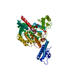 2bvmS C: citing same article ( S: Starting model for refinement |
|---|---|
| Similar structure data |


-Links
 PDBj
PDBj


- Assembly
| Deposited unit | 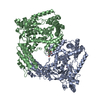
| ||||||||
|---|---|---|---|---|---|---|---|---|---|
| 1 | 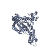
| ||||||||
| 2 | 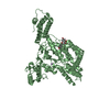
| ||||||||
| Unit cell |
|
-Components
| #1: Protein | Mass: 65356.988 Da / Num. of mol.: 2 / Fragment: residues 1-543 Source method: isolated from a genetically manipulated source Source: (gene. exp.) Clostridioides difficile (bacteria) / Gene: toxB, tcdB / Plasmid: pET28a+ / Production host: References: UniProt: P18177, Hydrolases; Acting on peptide bonds (peptidases); Cysteine endopeptidases #2: Chemical |   Mass: 54.938 Da / Num. of mol.: 2 / Source method: obtained synthetically / Formula: Mn Mass: 54.938 Da / Num. of mol.: 2 / Source method: obtained synthetically / Formula: Mn#3: Chemical | ChemComp-AGI / | 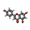  Mass: 270.237 Da / Num. of mol.: 1 / Source method: obtained synthetically / Formula: C15H10O5 Mass: 270.237 Da / Num. of mol.: 1 / Source method: obtained synthetically / Formula: C15H10O5 |
|---|
-Experimental details
-Experiment
| Experiment | Method: X-RAY DIFFRACTION / Number of used crystals: 1 |
|---|
- Sample preparation
| Crystal | Density Matthews: 3.3 Å3/Da / Density % sol: 62.7 % |
|---|---|
| Crystal grow | Temperature: 294 K / Method: vapor diffusion, hanging drop Details: 0.1 M HEPES pH 7.5-8.5, 0.2 M Mg(CH3CHOO)2, PEG 3350 5-25% |
-Data collection
| Diffraction | Mean temperature: 100 K |
|---|---|
| Diffraction source | Source: SYNCHROTRON / Site: APS / Beamline: 21-ID-G / Wavelength: 0.97856 Å |
| Detector | Type: RAYONIX MX-300 / Detector: CCD / Date: Apr 20, 2016 |
| Radiation | Protocol: SINGLE WAVELENGTH / Monochromatic (M) / Laue (L): M / Scattering type: x-ray |
| Radiation wavelength | Wavelength: 0.97856 Å / Relative weight: 1 |
| Reflection | Resolution: 2.75→44.437 Å / Num. obs: 45388 / % possible obs: 99.75 % / Redundancy: 6.6 % / CC1/2: 0.549 / Net I/σ(I): 21.33 |
- Processing
| Software |
| ||||||||||||||||||||||||||||||||||||||||||||||||||||||||||||||||||||||||||||||||||||||||||||||||||||||||||||||||||||||||||||||||||||||||||||||||||||||||||||||||||||||||||||||||||||||||||||||||||||||||||||||||||||||||||||||||||||||||||||||||||||||||||||||||||||||||||||||||||||||||||||||||||||||||||||
|---|---|---|---|---|---|---|---|---|---|---|---|---|---|---|---|---|---|---|---|---|---|---|---|---|---|---|---|---|---|---|---|---|---|---|---|---|---|---|---|---|---|---|---|---|---|---|---|---|---|---|---|---|---|---|---|---|---|---|---|---|---|---|---|---|---|---|---|---|---|---|---|---|---|---|---|---|---|---|---|---|---|---|---|---|---|---|---|---|---|---|---|---|---|---|---|---|---|---|---|---|---|---|---|---|---|---|---|---|---|---|---|---|---|---|---|---|---|---|---|---|---|---|---|---|---|---|---|---|---|---|---|---|---|---|---|---|---|---|---|---|---|---|---|---|---|---|---|---|---|---|---|---|---|---|---|---|---|---|---|---|---|---|---|---|---|---|---|---|---|---|---|---|---|---|---|---|---|---|---|---|---|---|---|---|---|---|---|---|---|---|---|---|---|---|---|---|---|---|---|---|---|---|---|---|---|---|---|---|---|---|---|---|---|---|---|---|---|---|---|---|---|---|---|---|---|---|---|---|---|---|---|---|---|---|---|---|---|---|---|---|---|---|---|---|---|---|---|---|---|---|---|---|---|---|---|---|---|---|---|---|---|---|---|---|---|---|---|---|---|---|---|---|---|---|---|---|---|---|---|---|---|---|---|---|---|---|---|---|---|---|---|---|---|---|---|---|---|---|---|---|---|
| Refinement | Method to determine structure: MOLECULAR REPLACEMENT Starting model: 2BVM Resolution: 2.75→44.437 Å / SU ML: 0.44 / Cross valid method: FREE R-VALUE / σ(F): 1.33 / Phase error: 26.18 / Stereochemistry target values: ML
| ||||||||||||||||||||||||||||||||||||||||||||||||||||||||||||||||||||||||||||||||||||||||||||||||||||||||||||||||||||||||||||||||||||||||||||||||||||||||||||||||||||||||||||||||||||||||||||||||||||||||||||||||||||||||||||||||||||||||||||||||||||||||||||||||||||||||||||||||||||||||||||||||||||||||||||
| Solvent computation | Shrinkage radii: 0.9 Å / VDW probe radii: 1.11 Å / Solvent model: FLAT BULK SOLVENT MODEL | ||||||||||||||||||||||||||||||||||||||||||||||||||||||||||||||||||||||||||||||||||||||||||||||||||||||||||||||||||||||||||||||||||||||||||||||||||||||||||||||||||||||||||||||||||||||||||||||||||||||||||||||||||||||||||||||||||||||||||||||||||||||||||||||||||||||||||||||||||||||||||||||||||||||||||||
| Refinement step | Cycle: LAST / Resolution: 2.75→44.437 Å
| ||||||||||||||||||||||||||||||||||||||||||||||||||||||||||||||||||||||||||||||||||||||||||||||||||||||||||||||||||||||||||||||||||||||||||||||||||||||||||||||||||||||||||||||||||||||||||||||||||||||||||||||||||||||||||||||||||||||||||||||||||||||||||||||||||||||||||||||||||||||||||||||||||||||||||||
| Refine LS restraints |
| ||||||||||||||||||||||||||||||||||||||||||||||||||||||||||||||||||||||||||||||||||||||||||||||||||||||||||||||||||||||||||||||||||||||||||||||||||||||||||||||||||||||||||||||||||||||||||||||||||||||||||||||||||||||||||||||||||||||||||||||||||||||||||||||||||||||||||||||||||||||||||||||||||||||||||||
| LS refinement shell |
| ||||||||||||||||||||||||||||||||||||||||||||||||||||||||||||||||||||||||||||||||||||||||||||||||||||||||||||||||||||||||||||||||||||||||||||||||||||||||||||||||||||||||||||||||||||||||||||||||||||||||||||||||||||||||||||||||||||||||||||||||||||||||||||||||||||||||||||||||||||||||||||||||||||||||||||
| Refinement TLS params. | Method: refined / Refine-ID: X-RAY DIFFRACTION
| ||||||||||||||||||||||||||||||||||||||||||||||||||||||||||||||||||||||||||||||||||||||||||||||||||||||||||||||||||||||||||||||||||||||||||||||||||||||||||||||||||||||||||||||||||||||||||||||||||||||||||||||||||||||||||||||||||||||||||||||||||||||||||||||||||||||||||||||||||||||||||||||||||||||||||||
| Refinement TLS group |
|
