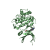
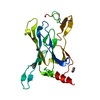
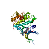
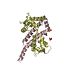
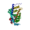
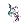
登録情報 データベース : PDB / ID : 5um4タイトル Crystal structure of the F255A mutant Kir3.1 cytoplasmic pore domain G protein-activated inward rectifier potassium channel 1 キーワード / 機能・相同性 分子機能 ドメイン・相同性 構成要素
/ / / / / / / / / / / / / / / / / / / / / / / / / / / / / / / / / / / / / / / / / / / / / / / 生物種 Anolis carolinensis (グリーンアノール)Mus musculus (ハツカネズミ)手法 / / / 解像度 : 2.5 Å データ登録者 Meinke, G. / Bohm, A. / Noujaim, S. 資金援助 組織 認可番号 国 National Institutes of Health/National Heart, Lung, and Blood Institute (NIH/NHLBI)
ジャーナル : To Be Published タイトル : Crystal structure of the F255A mutant Kir3.1 cytoplasmic pore domain著者 : Meinke, G. / Bohm, A. / Noujaim, S. 履歴 登録 2017年1月26日 登録サイト / 処理サイト 改定 1.0 2018年1月31日 Provider / タイプ 改定 1.1 2022年3月23日 Group / Database references / カテゴリ / pdbx_audit_supportItem / _database_2.pdbx_database_accession / _pdbx_audit_support.funding_organization改定 1.2 2023年10月4日 Group / Refinement descriptionカテゴリ / chem_comp_bond / pdbx_initial_refinement_model改定 1.3 2024年11月6日 Group カテゴリ / pdbx_modification_feature
すべて表示 表示を減らす
 ムービー
ムービー コントローラー


 データを開く
 基本情報
基本情報 要素
要素 キーワード
キーワード 機能・相同性情報
 Anolis carolinensis (グリーンアノール)
 X線回折 /
  データ登録者
 米国, 1件
  引用
引用 構造の表示
構造の表示 ダウンロードとリンク
 その他のダウンロード
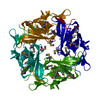


 PDBj
PDBj
 集合体
集合体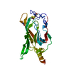

 分子量: 18.015 Da / 分子数: 27 / 由来タイプ: 天然 / 式: H2O
 試料調製
試料調製 解析
解析