 Movie
Movie Controller

[English] 日本語
 Yorodumi
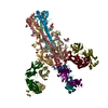
- PDB-5ug0: Human antibody H2897 in complex with influenza hemagglutinin H1 S... -
+ Open data

- Basic information
| Entry | Database: PDB / ID: 5ug0 | |||||||||
|---|---|---|---|---|---|---|---|---|---|---|
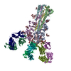
| Title | Human antibody H2897 in complex with influenza hemagglutinin H1 Solomon Islands/03/2006 | |||||||||
 Components Components |
| |||||||||
 Keywords Keywords | VIRAL PROTEIN/IMMUNE SYSTEM / Influenza HA / antibody / complex / VIRAL PROTEIN-IMMUNE SYSTEM complex | |||||||||
| Function / homology |  Function and homology information Function and homology informationimmunoglobulin complex / viral budding from plasma membrane / clathrin-dependent endocytosis of virus by host cell / adaptive immune response / host cell surface receptor binding / fusion of virus membrane with host plasma membrane / fusion of virus membrane with host endosome membrane / viral envelope / virion attachment to host cell / host cell plasma membrane ...immunoglobulin complex / viral budding from plasma membrane / clathrin-dependent endocytosis of virus by host cell / adaptive immune response / host cell surface receptor binding / fusion of virus membrane with host plasma membrane / fusion of virus membrane with host endosome membrane / viral envelope / virion attachment to host cell / host cell plasma membrane / virion membrane / extracellular region / membrane / plasma membrane Similarity search - Function | |||||||||
| Biological species |   Influenza A virus Influenza A virus Homo sapiens (human) Homo sapiens (human) | |||||||||
| Method |  X-RAY DIFFRACTION / SYNCHROTRON / MOLECULAR REPLACEMENT / molecular replacement / Resolution: 3.4 Å X-RAY DIFFRACTION / SYNCHROTRON / MOLECULAR REPLACEMENT / molecular replacement / Resolution: 3.4 Å | |||||||||
 Authors Authors | Raymond, D.D. / Caradonna, T. / Schmidt, A.G. / Harrison, S.C. | |||||||||
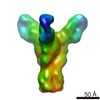
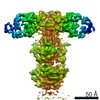
 Citation Citation | Journal: J Mol Biol / Year: 2017 Title: CryoEM Structure of an Influenza Virus Receptor-Binding Site Antibody-Antigen Interface. Authors: Yuhang Liu / Junhua Pan / Simon Jenni / Donald D Raymond / Tim Caradonna / Khoi T Do / Aaron G Schmidt / Stephen C Harrison / Nikolaus Grigorieff /  Abstract: Structure-based vaccine design depends on extensive structural analyses of antigen-antibody complexes.Single-particle electron cryomicroscopy (cryoEM) can circumvent some of the problems of x-ray ...Structure-based vaccine design depends on extensive structural analyses of antigen-antibody complexes.Single-particle electron cryomicroscopy (cryoEM) can circumvent some of the problems of x-ray crystallography as a pipeline for obtaining the required structures. We have examined the potential of single-particle cryoEM for determining the structure of influenza-virus hemagglutinin (HA):single-chain variable-domain fragment complexes, by studying a complex we failed to crystallize in pursuing an extended project on the human immune response to influenza vaccines.The result shows that a combination of cryoEM and molecular modeling can yield details of the antigen-antibody interface, although small variation in the twist of the rod-likeHA trimer limited the overall resolution to about 4.5Å.Comparison of principal 3D classes suggests ways to modify the HA trimer to overcome this limitation. A closely related antibody from the same donor did yield crystals when bound with the same HA, giving us an independent validation of the cryoEM results.The two structures also augment our understanding of receptor-binding site recognition by antibodies that neutralize a wide range of influenza-virus variants. | |||||||||
| History |
|
- Structure visualization
| Structure viewer | Molecule: MolmilJmol/JSmol |
|---|
- Downloads & links
-Download
| PDBx/mmCIF format | 5ug0.cif.gz | 201 KB | Display | PDBx/mmCIF format |
|---|---|---|---|---|
| PDB format | pdb5ug0.ent.gz | 156.8 KB | Display | PDB format |
| PDBx/mmJSON format | 5ug0.json.gz | Tree view | PDBx/mmJSON format | |
| Others |  Other downloads Other downloads |
-Validation report
| Arichive directory | https://data.pdbj.org/pub/pdb/validation_reports/ug/5ug0ftp://data.pdbj.org/pub/pdb/validation_reports/ug/5ug0 | HTTPS FTP |
|---|
-Related structure data
| Related structure data | 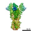 8561C  8562C 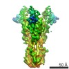 8563C 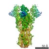 8564C  5ujzC  5uk0C  5uk1C  5uk2C  3sm5 S: Starting model for refinement C: citing same article ( |
|---|---|
| Similar structure data |


-Links
 PDBj
PDBj


- Assembly
| Deposited unit | 
| ||||||||
|---|---|---|---|---|---|---|---|---|---|
| 1 | 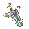
| ||||||||
| Unit cell |
|
-Components
-Hemagglutinin ... , 2 types, 2 molecules AB
| #1: Protein | Mass: 36509.902 Da / Num. of mol.: 1 / Fragment: UNP residues 18-343 Source method: isolated from a genetically manipulated source Source: (gene. exp.) Influenza A virus (A/Solomon Islands/3/2006(H1N1))Strain: A/Solomon Islands/3/2006(H1N1) / Gene: HA / Production host:  Trichoplusia ni (cabbage looper) / References: UniProt: A7UPX0 Trichoplusia ni (cabbage looper) / References: UniProt: A7UPX0 |
|---|---|
| #2: Protein | Mass: 20823.184 Da / Num. of mol.: 1 / Fragment: UNP residues 344-519 Source method: isolated from a genetically manipulated source Source: (gene. exp.) Influenza A virus (A/Solomon Islands/3/2006(H1N1))Strain: A/Solomon Islands/3/2006(H1N1) / Gene: HA / Production host: Trichoplusia ni (cabbage looper) / References: UniProt: A7UPX0 |
-Antibody , 2 types, 2 molecules CD
| #3: Antibody | Mass: 23530.148 Da / Num. of mol.: 1 Source method: isolated from a genetically manipulated source Source: (gene. exp.) Homo sapiens (human) / Gene: IGK@ / Production host: Homo sapiens (human) / References: UniProt: Q6PIL8 |
|---|---|
| #4: Antibody | Mass: 24537.402 Da / Num. of mol.: 1 Source method: isolated from a genetically manipulated source Source: (gene. exp.) Homo sapiens (human) / Gene: DKFZp686P15220 / Production host: Homo sapiens (human) / References: UniProt: Q6N089 |
-Sugars , 3 types, 8 molecules 
| #5: Polysaccharide | Source method: isolated from a genetically manipulated source #6: Polysaccharide | 2-acetamido-2-deoxy-beta-D-glucopyranose-(1-4)-2-acetamido-2-deoxy-beta-D-glucopyranose | Source method: isolated from a genetically manipulated source #7: Sugar | ChemComp-NAG /  Type: D-saccharide, beta linking / Mass: 221.208 Da / Num. of mol.: 5 Type: D-saccharide, beta linking / Mass: 221.208 Da / Num. of mol.: 5Source method: isolated from a genetically manipulated source Formula: C8H15NO6 |
|---|
-Details
| Has protein modification | Y |
|---|
-Experimental details
-Experiment
| Experiment | Method: X-RAY DIFFRACTION / Number of used crystals: 1 |
|---|
- Sample preparation
| Crystal | Density Matthews: 5.68 Å3/Da / Density % sol: 78.35 % |
|---|---|
| Crystal grow | Temperature: 293.15 K / Method: vapor diffusion, hanging drop / Details: 10% w/v PEG8000, 100 mM HEPES |
-Data collection
| Diffraction | Mean temperature: 293.15 K | ||||||||||||||||||||||||||||||||||||||||||||||||||||||||||||||||||||||
|---|---|---|---|---|---|---|---|---|---|---|---|---|---|---|---|---|---|---|---|---|---|---|---|---|---|---|---|---|---|---|---|---|---|---|---|---|---|---|---|---|---|---|---|---|---|---|---|---|---|---|---|---|---|---|---|---|---|---|---|---|---|---|---|---|---|---|---|---|---|---|---|
| Diffraction source | Source: SYNCHROTRON / Site: APS / Beamline: 24-ID-E / Wavelength: 0.97923 Å | ||||||||||||||||||||||||||||||||||||||||||||||||||||||||||||||||||||||
| Detector | Type: ADSC QUANTUM 315 / Detector: CCD / Date: Aug 5, 2015 | ||||||||||||||||||||||||||||||||||||||||||||||||||||||||||||||||||||||
| Radiation | Monochromator: Cryogenically-cooled single crystal Si(220) / Protocol: SINGLE WAVELENGTH / Monochromatic (M) / Laue (L): M / Scattering type: x-ray | ||||||||||||||||||||||||||||||||||||||||||||||||||||||||||||||||||||||
| Radiation wavelength | Wavelength: 0.97923 Å / Relative weight: 1 | ||||||||||||||||||||||||||||||||||||||||||||||||||||||||||||||||||||||
| Reflection | Resolution: 3.4→46.41 Å / Num. obs: 32426 / % possible obs: 98.6 % / Observed criterion σ(I): -3 / Redundancy: 3.672 % / Biso Wilson estimate: 116.33 Å2 / CC1/2: 0.987 / Rmerge(I) obs: 0.274 / Rrim(I) all: 0.322 / Χ2: 1.007 / Net I/σ(I): 5.75 / Num. measured all: 119057 / Scaling rejects: 64 | ||||||||||||||||||||||||||||||||||||||||||||||||||||||||||||||||||||||
| Reflection shell |
|
-Phasing
| Phasing | Method: molecular replacement |
|---|
- Processing
| Software |
| ||||||||||||||||||||||||||||||||||||||||||||||||||||||||||||||||||||||||||||||||||||||||||||||||||||||||||||
|---|---|---|---|---|---|---|---|---|---|---|---|---|---|---|---|---|---|---|---|---|---|---|---|---|---|---|---|---|---|---|---|---|---|---|---|---|---|---|---|---|---|---|---|---|---|---|---|---|---|---|---|---|---|---|---|---|---|---|---|---|---|---|---|---|---|---|---|---|---|---|---|---|---|---|---|---|---|---|---|---|---|---|---|---|---|---|---|---|---|---|---|---|---|---|---|---|---|---|---|---|---|---|---|---|---|---|---|---|---|
| Refinement | Method to determine structure: MOLECULAR REPLACEMENT Starting model: PDB entry 3SM5 3sm5 Resolution: 3.4→46.41 Å / Cor.coef. Fo:Fc: 0.865 / Cor.coef. Fo:Fc free: 0.848 / Rfactor Rfree error: 0 / SU R Cruickshank DPI: 1.344 / Cross valid method: THROUGHOUT / σ(F): 0 / SU R Blow DPI: 1.218 / SU Rfree Blow DPI: 0.442 / SU Rfree Cruickshank DPI: 0.453
| ||||||||||||||||||||||||||||||||||||||||||||||||||||||||||||||||||||||||||||||||||||||||||||||||||||||||||||
| Displacement parameters | Biso max: 296.08 Å2 / Biso mean: 158.46 Å2 / Biso min: 98.19 Å2
| ||||||||||||||||||||||||||||||||||||||||||||||||||||||||||||||||||||||||||||||||||||||||||||||||||||||||||||
| Refine analyze | Luzzati coordinate error obs: 0.87 Å | ||||||||||||||||||||||||||||||||||||||||||||||||||||||||||||||||||||||||||||||||||||||||||||||||||||||||||||
| Refinement step | Cycle: final / Resolution: 3.4→46.41 Å
| ||||||||||||||||||||||||||||||||||||||||||||||||||||||||||||||||||||||||||||||||||||||||||||||||||||||||||||
| Refine LS restraints |
| ||||||||||||||||||||||||||||||||||||||||||||||||||||||||||||||||||||||||||||||||||||||||||||||||||||||||||||
| LS refinement shell | Resolution: 3.4→3.51 Å / Rfactor Rfree error: 0 / Total num. of bins used: 16
|
