 Movie
Movie Controller

[English] 日本語
 Yorodumi
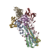
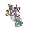
- PDB-5uk0: CryoEM structure of an influenza virus receptor-binding site anti... -
+ Open data

- Basic information
| Entry | Database: PDB / ID: 5uk0 | |||||||||
|---|---|---|---|---|---|---|---|---|---|---|
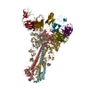
| Title | CryoEM structure of an influenza virus receptor-binding site antibody-antigen interface - Class 2 | |||||||||
 Components Components |
| |||||||||
 Keywords Keywords | VIRAL PROTEIN/IMMUNE SYSTEM / viral glycoprotein / hemagglutinin / antibody fragment / VIRAL PROTEIN-IMMUNE SYSTEM complex | |||||||||
| Function / homology |  Function and homology information Function and homology informationviral budding from plasma membrane / clathrin-dependent endocytosis of virus by host cell / host cell surface receptor binding / fusion of virus membrane with host plasma membrane / fusion of virus membrane with host endosome membrane / viral envelope / virion attachment to host cell / host cell plasma membrane / virion membrane / membrane Similarity search - Function | |||||||||
| Biological species |   Influenza A virus Influenza A virus Homo sapiens (human) Homo sapiens (human) | |||||||||
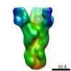
| Method | ELECTRON MICROSCOPY / single particle reconstruction / cryo EM / Resolution: 4.8 Å | |||||||||
 Authors Authors | Liu, Y. / Pan, J. / Caradonna, T. / Jenni, S. / Raymond, D.D. / Schmidt, A.G. / Harrison, S.C. / Grigorieff, N. | |||||||||
| Funding support |  United States, 1items United States, 1items
| |||||||||
 Citation Citation | Journal: J Mol Biol / Year: 2017 Title: CryoEM Structure of an Influenza Virus Receptor-Binding Site Antibody-Antigen Interface. Authors: Yuhang Liu / Junhua Pan / Simon Jenni / Donald D Raymond / Tim Caradonna / Khoi T Do / Aaron G Schmidt / Stephen C Harrison / Nikolaus Grigorieff / Abstract: Structure-based vaccine design depends on extensive structural analyses of antigen-antibody complexes.Single-particle electron cryomicroscopy (cryoEM) can circumvent some of the problems of x-ray ...Structure-based vaccine design depends on extensive structural analyses of antigen-antibody complexes.Single-particle electron cryomicroscopy (cryoEM) can circumvent some of the problems of x-ray crystallography as a pipeline for obtaining the required structures. We have examined the potential of single-particle cryoEM for determining the structure of influenza-virus hemagglutinin (HA):single-chain variable-domain fragment complexes, by studying a complex we failed to crystallize in pursuing an extended project on the human immune response to influenza vaccines.The result shows that a combination of cryoEM and molecular modeling can yield details of the antigen-antibody interface, although small variation in the twist of the rod-likeHA trimer limited the overall resolution to about 4.5Å.Comparison of principal 3D classes suggests ways to modify the HA trimer to overcome this limitation. A closely related antibody from the same donor did yield crystals when bound with the same HA, giving us an independent validation of the cryoEM results.The two structures also augment our understanding of receptor-binding site recognition by antibodies that neutralize a wide range of influenza-virus variants. | |||||||||
| History |
|
- Structure visualization
| Movie |
Movie viewer |
|---|---|
| Structure viewer | Molecule: MolmilJmol/JSmol |
- Downloads & links
-Download
| PDBx/mmCIF format | 5uk0.cif.gz | 713.4 KB | Display | PDBx/mmCIF format |
|---|---|---|---|---|
| PDB format | pdb5uk0.ent.gz | 601.4 KB | Display | PDB format |
| PDBx/mmJSON format | 5uk0.json.gz | Tree view | PDBx/mmJSON format | |
| Others |  Other downloads Other downloads |
-Validation report
| Arichive directory | https://data.pdbj.org/pub/pdb/validation_reports/uk/5uk0ftp://data.pdbj.org/pub/pdb/validation_reports/uk/5uk0 | HTTPS FTP |
|---|
-Related structure data
| Related structure data |  8562MC 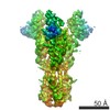 8561C 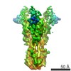 8563C 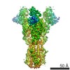 8564C 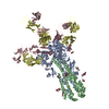 5ug0C 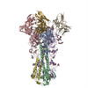 5ujzC 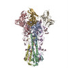 5uk1C  5uk2C M: map data used to model this data C: citing same article ( |
|---|---|
| Similar structure data |

-Links
 PDBj
PDBj


- Assembly
| Deposited unit | 
|
|---|---|
| 1 |
|
-Components
-Hemagglutinin ... , 2 types, 6 molecules ACEBDF
| #1: Protein | Mass: 36024.344 Da / Num. of mol.: 3 / Fragment: UNP residues 18-339 Source method: isolated from a genetically manipulated source Source: (gene. exp.) Influenza A virus (A/Solomon Islands/3/2006(H1N1))Strain: A/Solomon Islands/3/2006(H1N1) / Gene: HA / Plasmid: pFastBac / Details (production host): PFASTBAC / Cell line (production host): HI-5 / Production host:  Trichoplusia ni (cabbage looper) / References: UniProt: A7UPX0 Trichoplusia ni (cabbage looper) / References: UniProt: A7UPX0#2: Protein | Mass: 19841.041 Da / Num. of mol.: 3 / Fragment: UNP residues 344-516 Source method: isolated from a genetically manipulated source Source: (gene. exp.) Influenza A virus / Strain: A/Solomon Islands/3/2006(H1N1) / Gene: HA / Plasmid: pFASTBAC / Cell line (production host): HI-5 / Production host: Trichoplusia ni (cabbage looper) / References: UniProt: A7UPX0 |
|---|
-Antibody , 1 types, 3 molecules GHI
| #3: Antibody | Mass: 27629.385 Da / Num. of mol.: 3 Source method: isolated from a genetically manipulated source Source: (gene. exp.) Homo sapiens (human) / Plasmid: pVRC / Cell line (production host): HEK293T / Production host: Homo sapiens (human) |
|---|
-Sugars , 3 types, 24 molecules 
| #4: Polysaccharide | Source method: isolated from a genetically manipulated source #5: Polysaccharide | Source method: isolated from a genetically manipulated source #6: Sugar | ChemComp-NAG /  Type: D-saccharide, beta linking / Mass: 221.208 Da / Num. of mol.: 18 Type: D-saccharide, beta linking / Mass: 221.208 Da / Num. of mol.: 18Source method: isolated from a genetically manipulated source Formula: C8H15NO6 |
|---|
-Details
| Has protein modification | Y |
|---|
-Experimental details
-Experiment
| Experiment | Method: ELECTRON MICROSCOPY |
|---|---|
| EM experiment | Aggregation state: PARTICLE / 3D reconstruction method: single particle reconstruction |
- Sample preparation
| Component | Name: Influenza-virus hemagglutinin H1 complexed with K1915 single-chain variable-domain fragment Type: COMPLEX Details: The complex consists of three hemagglutinin head domains bound to three single-chain variable-domain fragments. Entity ID: #1-#3 / Source: RECOMBINANT |
|---|---|
| Molecular weight | Value: 0.32 MDa / Experimental value: NO |
| Source (natural) | Organism: Influenza A virus (A/Solomon Islands/3/2006(H1N1)) |
| Source (recombinant) | Organism: Trichoplusia ni (cabbage looper) / Cell: Hi-5 |
| Buffer solution | pH: 8 Details: Beta-octylglucoside was added to a final concentration of 0.07% w/v to induce more variable particle orientations. |
| Specimen | Conc.: 0.1 mg/ml / Embedding applied: NO / Shadowing applied: NO / Staining applied: NO / Vitrification applied: YES |
| Specimen support | Grid material: COPPER / Grid mesh size: 200 divisions/in. / Grid type: Quantifoil R1.2/1.3 |
| Vitrification | Instrument: FEI VITROBOT MARK I / Cryogen name: ETHANE / Humidity: 90 % / Chamber temperature: 298 K |
- Electron microscopy imaging
| Experimental equipment |  Model: Titan Krios / Image courtesy: FEI Company |
|---|---|
| Microscopy | Model: FEI TITAN KRIOS |
| Electron gun | Electron source:  FIELD EMISSION GUN / Accelerating voltage: 300 kV / Illumination mode: FLOOD BEAM FIELD EMISSION GUN / Accelerating voltage: 300 kV / Illumination mode: FLOOD BEAM |
| Electron lens | Mode: BRIGHT FIELD / Nominal magnification: 18000 X / Calibrated magnification: 30444 X / Calibrated defocus min: 1000 nm / Calibrated defocus max: 9000 nm / Cs: 2.7 mm / Alignment procedure: BASIC |
| Specimen holder | Cryogen: NITROGEN / Specimen holder model: FEI TITAN KRIOS AUTOGRID HOLDER / Temperature (max): 70 K / Temperature (min): 70 K |
| Image recording | Average exposure time: 13 sec. / Electron dose: 40 e/Å2 / Detector mode: SUPER-RESOLUTION / Film or detector model: GATAN K2 SUMMIT (4k x 4k) / Num. of grids imaged: 1 / Num. of real images: 10281 Details: The exposure rate was 8 electrons/physical pixel/second. |
| Image scans | Sampling size: 2.5 µm / Width: 7676 / Height: 7420 / Movie frames/image: 38 / Used frames/image: 1-38 |
- Processing
| EM software |
| |||||||||||||||||||||||||||||||||||||||||||||
|---|---|---|---|---|---|---|---|---|---|---|---|---|---|---|---|---|---|---|---|---|---|---|---|---|---|---|---|---|---|---|---|---|---|---|---|---|---|---|---|---|---|---|---|---|---|---|
| Image processing | Details: The particle images were normalized to have constant variance and zero average. Movies were processed using Unblur. | |||||||||||||||||||||||||||||||||||||||||||||
| CTF correction | Type: PHASE FLIPPING AND AMPLITUDE CORRECTION | |||||||||||||||||||||||||||||||||||||||||||||
| Particle selection | Num. of particles selected: 252130 | |||||||||||||||||||||||||||||||||||||||||||||
| 3D reconstruction | Resolution: 4.8 Å / Resolution method: FSC 0.143 CUT-OFF / Num. of particles: 142314 / Algorithm: FOURIER SPACE Details: Refinement and classification were limited to 10 Angstrom resolution. Num. of class averages: 3 / Symmetry type: POINT | |||||||||||||||||||||||||||||||||||||||||||||
| Atomic model building | Protocol: OTHER / Space: REAL / Target criteria: Correlation coefficient Details: Domains were initially placed manually using Chimera. The HA trimer was from PDB ID 5UGY. Fab was initially obtained using MODELLER, with PDB ID 4K8R as template. The structure was refined using PHENIX. |
