[English] 日本語
Yorodumi
- PDB-5uf5: Structure of the effector protein SidK (lpg0968) from Legionella ... -

+
Open data


ID or keywords:

Loading...

-
Basic information

Entry
Database: PDB / ID: 5uf5
TitleStructure of the effector protein SidK (lpg0968) from Legionella pneumophila (domain-swapped dimer)
Componentseffector protein SidK
KeywordsPROTEIN BINDING / translocated effector / V-ATPase binding / all-alpha-helical
Function / homology: / Type IV secretion protein Dot
Function and homology information
Biological speciesLegionella pneumophila (bacteria)
MethodX-RAY DIFFRACTION / SYNCHROTRON / SAD / Resolution: 2.4 Å
AuthorsBeyrakhova, K. / Xu, C. / Boniecki, M.T. / Cygler, M.
Funding support Canada, 1items
OrganizationGrant numberCountry
Canadian Institutes of Health Research (CIHR)MOP-48370 Canada
Citation
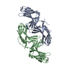
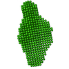
Journal: PLoS Pathog / Year: 2017
Title: Molecular basis for the binding and modulation of V-ATPase by a bacterial effector protein.
Authors: Jianhua Zhao / Ksenia Beyrakhova / Yao Liu / Claudia P Alvarez / Stephanie A Bueler / Li Xu / Caishuang Xu / Michal T Boniecki / Voula Kanelis / Zhao-Qing Luo / Miroslaw Cygler / John L Rubinstein /
Abstract: Intracellular pathogenic bacteria evade the immune response by replicating within host cells. Legionella pneumophila, the causative agent of Legionnaires' Disease, makes use of numerous effector ...Intracellular pathogenic bacteria evade the immune response by replicating within host cells. Legionella pneumophila, the causative agent of Legionnaires' Disease, makes use of numerous effector proteins to construct a niche supportive of its replication within phagocytic cells. The L. pneumophila effector SidK was identified in a screen for proteins that reduce the activity of the proton pumping vacuolar-type ATPases (V-ATPases) when expressed in the yeast Saccharomyces cerevisae. SidK is secreted by L. pneumophila in the early stages of infection and by binding to and inhibiting the V-ATPase, SidK reduces phagosomal acidification and promotes survival of the bacterium inside macrophages. We determined crystal structures of the N-terminal region of SidK at 2.3 Å resolution and used single particle electron cryomicroscopy (cryo-EM) to determine structures of V-ATPase:SidK complexes at ~6.8 Å resolution. SidK is a flexible and elongated protein composed of an α-helical region that interacts with subunit A of the V-ATPase and a second region of unknown function that is flexibly-tethered to the first. SidK binds V-ATPase strongly by interacting via two α-helical bundles at its N terminus with subunit A. In vitro activity assays show that SidK does not inhibit the V-ATPase completely, but reduces its activity by ~40%, consistent with the partial V-ATPase deficiency phenotype its expression causes in yeast. The cryo-EM analysis shows that SidK reduces the flexibility of the A-subunit that is in the 'open' conformation. Fluorescence experiments indicate that SidK binding decreases the affinity of V-ATPase for a fluorescent analogue of ATP. Together, these results reveal the structural basis for the fine-tuning of V-ATPase activity by SidK.
History
DepositionJan 3, 2017Deposition site: RCSB / Processing site: RCSB
Revision 1.0May 10, 2017Provider: repository / Type: Initial release
Revision 1.1Jun 21, 2017Group: Database references / Category: citation / citation_author
Item: _citation.journal_abbrev / _citation.journal_id_CSD ..._citation.journal_abbrev / _citation.journal_id_CSD / _citation.journal_id_ISSN / _citation.journal_volume / _citation.page_first / _citation.page_last / _citation.pdbx_database_id_DOI / _citation.pdbx_database_id_PubMed / _citation.title / _citation.year / _citation_author.name
Revision 1.2Sep 20, 2017Group: Author supporting evidence / Category: pdbx_audit_support / Item: _pdbx_audit_support.funding_organization
Revision 1.3Jan 8, 2020Group: Author supporting evidence / Category: pdbx_audit_support / Item: _pdbx_audit_support.funding_organization
Revision 1.4Mar 6, 2024Group: Data collection / Database references / Category: chem_comp_atom / chem_comp_bond / database_2
Item: _database_2.pdbx_DOI / _database_2.pdbx_database_accession

-
Structure visualization

Structure viewerMolecule:
MolmilJmol/JSmol

Downloads & links

-
Assembly

Deposited unit
A: effector protein SidK
B: effector protein SidK
hetero molecules


Theoretical massNumber of molelcules
Total (without water)61,3663
Polymers61,2742
Non-polymers921
Water1,56787
1


  • Idetical with deposited unit
  • defined by author&software
TypeNameSymmetry operationNumber
identity operation1_555x,y,z1
Buried area3530 Å2
ΔGint-18 kcal/mol
Surface area24690 Å2
MethodPISA
Unit cell
Length a, b, c (Å)162.080, 41.140, 94.150
Angle α, β, γ (deg.)90.00, 108.39, 90.00
Int Tables number5
Space group name H-MC121

-
Components

#1: Protein effector protein SidK


Mass: 30637.191 Da / Num. of mol.: 2
Source method: isolated from a genetically manipulated source
Source: (gene. exp.) Legionella pneumophila (bacteria) / Gene: lpg_0968 / Production host: Escherichia coli (E. coli) / References: UniProt: G8UUS6, UniProt: Q5ZWW6*PLUS
#2: Chemical ChemComp-GOL / GLYCEROL / GLYCERIN / PROPANE-1,2,3-TRIOL


Mass: 92.094 Da / Num. of mol.: 1 / Source method: obtained synthetically / Formula: C3H8O3
#3: Water ChemComp-HOH / water


Mass: 18.015 Da / Num. of mol.: 87 / Source method: isolated from a natural source / Formula: H2O

-
Experimental details

-
Experiment

ExperimentMethod: X-RAY DIFFRACTION / Number of used crystals: 1

-
Sample preparation

CrystalDensity Matthews: 2.42 Å3/Da / Density % sol: 49 % / Description: plates
Crystal growTemperature: 293 K / Method: vapor diffusion, hanging drop / pH: 6.5
Details: 0.1 M sodium cacodylate trihydrate pH 6.5, 0.2 M magnesium acetate tetrahydrate, 20% (w/v) polyethylene glycol 8,000

-
Data collection

DiffractionMean temperature: 100 K
Diffraction sourceSource: SYNCHROTRON / Site: CLSI / Beamline: 08ID-1 / Wavelength: 0.9794 Å
DetectorType: RAYONIX MX-300 / Detector: CCD / Date: Mar 8, 2016
RadiationProtocol: SINGLE WAVELENGTH / Monochromatic (M) / Laue (L): M / Scattering type: x-ray
Radiation wavelengthWavelength: 0.9794 Å / Relative weight: 1
ReflectionResolution: 2.4→45.34 Å / Num. obs: 23542 / % possible obs: 99.9 % / Redundancy: 6.2 % / CC1/2: 0.998 / Rmerge(I) obs: 0.125 / Rsym value: 0.115 / Net I/σ(I): 13.4
Reflection shellResolution: 2.4→2.46 Å / Redundancy: 6.3 % / Rmerge(I) obs: 1.05 / Mean I/σ(I) obs: 1.98 / CC1/2: 0.7 / % possible all: 99.8

-
Processing

Software
NameVersionClassification
PHENIX(1.10.1_2155: ???)refinement
XDSdata reduction
XDSdata scaling
SHELXDphasing
RefinementMethod to determine structure: SAD / Resolution: 2.4→45.336 Å / SU ML: 0.33 / Cross valid method: FREE R-VALUE / σ(F): 1.35 / Phase error: 25.14
RfactorNum. reflection% reflection
Rfree0.2296 1178 5.01 %
Rwork0.1954 --
obs0.1972 23532 99.94 %
Solvent computationShrinkage radii: 0.9 Å / VDW probe radii: 1.11 Å
Refinement stepCycle: LAST / Resolution: 2.4→45.336 Å
ProteinNucleic acidLigandSolventTotal
Num. atoms3939 0 6 87 4032
Refine LS restraints
Refine-IDTypeDev idealNumber
X-RAY DIFFRACTIONf_bond_d0.0024019
X-RAY DIFFRACTIONf_angle_d0.4545452
X-RAY DIFFRACTIONf_dihedral_angle_d12.0692469
X-RAY DIFFRACTIONf_chiral_restr0.035626
X-RAY DIFFRACTIONf_plane_restr0.003708
LS refinement shell
Resolution (Å)Rfactor RfreeNum. reflection RfreeRfactor RworkNum. reflection RworkRefine-ID% reflection obs (%)
2.4-2.50920.33141450.26852743X-RAY DIFFRACTION100
2.5092-2.64150.32091450.24172756X-RAY DIFFRACTION100
2.6415-2.80690.27661460.23172775X-RAY DIFFRACTION100
2.8069-3.02360.27181460.21952769X-RAY DIFFRACTION100
3.0236-3.32780.28611460.21842788X-RAY DIFFRACTION100
3.3278-3.80920.19891480.18852800X-RAY DIFFRACTION100
3.8092-4.79830.19371480.16212814X-RAY DIFFRACTION100
4.7983-45.3440.19931540.18152909X-RAY DIFFRACTION100
Refinement TLS params.

Method: refined / Refine-ID: X-RAY DIFFRACTION

IDL112)L122)L132)L222)L232)L332)S11 (Å °)S12 (Å °)S13 (Å °)S21 (Å °)S22 (Å °)S23 (Å °)S31 (Å °)S32 (Å °)S33 (Å °)T112)T122)T132)T222)T232)T332)Origin x (Å)Origin y (Å)Origin z (Å)
13.7878-3.01280.01893.1031-1.10223.22330.20590.34430.2056-0.2686-0.1809-0.0846-0.0216-0.0136-0.03930.3327-0.03840.02050.3249-0.02880.291132.0136-22.28977.6775
23.4346-2.42011.10364.7726-0.79493.7155-0.05170.01830.5186-0.204-0.09490.0705-0.6128-0.81130.08730.43760.0957-0.02950.4407-0.07130.44067.2697-3.901520.7917
31.80750.619-1.68464.0993.05484.96460.2675-0.07810.5325-1.7590.04681.5179-1.4714-1.2982-0.30651.07440.456-0.38211.12-0.34251.0948-8.75055.010712.5744
42.6907-0.92981.53161.1475-0.68362.6846-0.0002-0.06190.02680.118-0.06650.0239-0.01190.01910.07210.4004-0.03620.0690.2697-0.0080.327929.6809-20.93440.4775
54.3844-1.62521.45012.7144-0.2452.7978-0.01230.0091-0.06360.3880.2736-0.64280.13840.5714-0.18270.39720.0954-0.0420.4989-0.10710.462155.9721-36.859628.4589
66.33811.537-0.70034.7281.24730.54320.961-0.1022-1.1631.1814-0.2174-0.56251.34971.0011-0.74821.0030.2215-0.47950.7252-0.23571.149569.1887-50.195736.9035
Refinement TLS group
IDRefine-IDRefine TLS-IDSelection details
1X-RAY DIFFRACTION1chain 'A' and (resid 16 through 122)
2X-RAY DIFFRACTION2chain 'A' and (resid 123 through 256 )
3X-RAY DIFFRACTION3chain 'A' and (resid 257 through 273 )
4X-RAY DIFFRACTION4chain 'B' and (resid 16 through 122 )
5X-RAY DIFFRACTION5chain 'B' and (resid 123 through 256 )
6X-RAY DIFFRACTION6chain 'B' and (resid 257 through 273 )

+
About Yorodumi

-
News

-
Feb 9, 2022. New format data for meta-information of EMDB entries

New format data for meta-information of EMDB entries

  • Version 3 of the EMDB header file is now the official format.
  • The previous official version 1.9 will be removed from the archive.

Related info.:EMDB header

External links:wwPDB to switch to version 3 of the EMDB data model

-
Aug 12, 2020. Covid-19 info

Covid-19 info

URL: https://pdbjlvh1.pdbj.org/emnavi/covid19.php

New page: Covid-19 featured information page in EM Navigator.

Related info.:Covid-19 info / Mar 5, 2020. Novel coronavirus structure data

+
Mar 5, 2020. Novel coronavirus structure data

Novel coronavirus structure data

Related info.:Yorodumi Speices / Aug 12, 2020. Covid-19 info

External links:COVID-19 featured content - PDBj / Molecule of the Month (242):Coronavirus Proteases

+
Jan 31, 2019. EMDB accession codes are about to change! (news from PDBe EMDB page)

EMDB accession codes are about to change! (news from PDBe EMDB page)

  • The allocation of 4 digits for EMDB accession codes will soon come to an end. Whilst these codes will remain in use, new EMDB accession codes will include an additional digit and will expand incrementally as the available range of codes is exhausted. The current 4-digit format prefixed with “EMD-” (i.e. EMD-XXXX) will advance to a 5-digit format (i.e. EMD-XXXXX), and so on. It is currently estimated that the 4-digit codes will be depleted around Spring 2019, at which point the 5-digit format will come into force.
  • The EM Navigator/Yorodumi systems omit the EMD- prefix.

Related info.:Q: What is EMD? / ID/Accession-code notation in Yorodumi/EM Navigator

External links:EMDB Accession Codes are Changing Soon! / Contact to PDBj

+
Jul 12, 2017. Major update of PDB

Major update of PDB

  • wwPDB released updated PDB data conforming to the new PDBx/mmCIF dictionary.
  • This is a major update changing the version number from 4 to 5, and with Remediation, in which all the entries are updated.
  • In this update, many items about electron microscopy experimental information are reorganized (e.g. em_software).
  • Now, EM Navigator and Yorodumi are based on the updated data.

External links:wwPDB Remediation / Enriched Model Files Conforming to OneDep Data Standards Now Available in the PDB FTP Archive

-
Yorodumi

Thousand views of thousand structures

  • Yorodumi is a browser for structure data from EMDB, PDB, SASBDB, etc.
  • This page is also the successor to EM Navigator detail page, and also detail information page/front-end page for Omokage search.
  • The word "yorodu" (or yorozu) is an old Japanese word meaning "ten thousand". "mi" (miru) is to see.

Related info.:EMDB / PDB / SASBDB / Comparison of 3 databanks / Yorodumi Search / Aug 31, 2016. New EM Navigator & Yorodumi / Yorodumi Papers / Jmol/JSmol / Function and homology information / Changes in new EM Navigator and Yorodumi

Read more