 Movie
Movie Controller

[English] 日本語
 Yorodumi
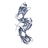
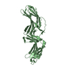
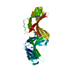
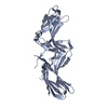
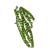
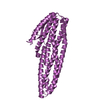
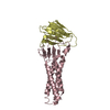
- PDB-3lh9: Crystal structure of mouse VPS26B(L197S/R199E) in spacegroup P41 21 2 -
+ Open data

- Basic information
| Entry | Database: PDB / ID: 3lh9 | ||||||
|---|---|---|---|---|---|---|---|
| Title | Crystal structure of mouse VPS26B(L197S/R199E) in spacegroup P41 21 2 | ||||||
 Components Components | Vacuolar protein sorting-associated protein 26B | ||||||
 Keywords Keywords | PROTEIN TRANSPORT / arrestin / fibronectin / Membrane / Transport | ||||||
| Function / homology |  Function and homology information Function and homology informationpostsynaptic early endosome / postsynaptic recycling endosome / retromer complex / retrograde transport, endosome to Golgi / regulation of neurotransmitter receptor localization to postsynaptic specialization membrane / phagocytic vesicle / intracellular protein transport / cellular response to type II interferon / late endosome / early endosome ...postsynaptic early endosome / postsynaptic recycling endosome / retromer complex / retrograde transport, endosome to Golgi / regulation of neurotransmitter receptor localization to postsynaptic specialization membrane / phagocytic vesicle / intracellular protein transport / cellular response to type II interferon / late endosome / early endosome / endosome / glutamatergic synapse / cytosol Similarity search - Function | ||||||
| Biological species |  | ||||||
| Method |  X-RAY DIFFRACTION / SYNCHROTRON / MOLECULAR REPLACEMENT / Resolution: 2.4 Å X-RAY DIFFRACTION / SYNCHROTRON / MOLECULAR REPLACEMENT / Resolution: 2.4 Å | ||||||
 Authors Authors | Collins, B. / Shaw, D. / Norwood, S. | ||||||
 Citation Citation | Journal: Traffic / Year: 2011 Title: Assembly and solution structure of the core retromer protein complex. Authors: Norwood, S.J. / Shaw, D.J. / Cowieson, N.P. / Owen, D.J. / Teasdale, R.D. / Collins, B.M. | ||||||
| History |
|
- Structure visualization
| Structure viewer | Molecule: MolmilJmol/JSmol |
|---|
- Downloads & links
-Download
| PDBx/mmCIF format | 3lh9.cif.gz | 138.2 KB | Display | PDBx/mmCIF format |
|---|---|---|---|---|
| PDB format | pdb3lh9.ent.gz | 107.7 KB | Display | PDB format |
| PDBx/mmJSON format | 3lh9.json.gz | Tree view | PDBx/mmJSON format | |
| Others |  Other downloads Other downloads |
-Validation report
| Arichive directory | https://data.pdbj.org/pub/pdb/validation_reports/lh/3lh9ftp://data.pdbj.org/pub/pdb/validation_reports/lh/3lh9 | HTTPS FTP |
|---|
-Related structure data
| Related structure data |  3lh8C  3lhaC  2r51S S: Starting model for refinement C: citing same article ( |
|---|---|
| Similar structure data |


-Links
 PDBj
PDBj- Assembly
| Deposited unit | 
| ||||||||
|---|---|---|---|---|---|---|---|---|---|
| 1 | 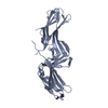
| ||||||||
| 2 | 
| ||||||||
| Unit cell |
|
-Components
| #1: Protein | Mass: 39648.973 Da / Num. of mol.: 2 / Fragment: UNP residues 7-336 / Mutation: L197S,R199E Source method: isolated from a genetically manipulated source Source: (gene. exp.)  #2: Water | ChemComp-HOH / |  Mass: 18.015 Da / Num. of mol.: 308 / Source method: isolated from a natural source / Formula: H2O Mass: 18.015 Da / Num. of mol.: 308 / Source method: isolated from a natural source / Formula: H2O |
|---|
-Experimental details
-Experiment
| Experiment | Method: X-RAY DIFFRACTION / Number of used crystals: 1 |
|---|
- Sample preparation
| Crystal | Density Matthews: 4.74 Å3/Da / Density % sol: 74.03 % |
|---|---|
| Crystal grow | Temperature: 289 K / Method: vapor diffusion, hanging drop / pH: 7.5 Details: 2M sodium formate, pH 7.5, VAPOR DIFFUSION, HANGING DROP, temperature 289K |
-Data collection
| Diffraction | Mean temperature: 100 K |
|---|---|
| Diffraction source | Source: SYNCHROTRON / Site: Australian Synchrotron  / Beamline: MX1 / Wavelength: 0.95364 Å / Beamline: MX1 / Wavelength: 0.95364 Å |
| Detector | Detector: CCD / Date: Nov 27, 2009 |
| Radiation | Protocol: SINGLE WAVELENGTH / Monochromatic (M) / Laue (L): M / Scattering type: x-ray |
| Radiation wavelength | Wavelength: 0.95364 Å / Relative weight: 1 |
| Reflection | Resolution: 2.4→45.2 Å / Num. obs: 60249 / % possible obs: 100 % / Redundancy: 8.6 % / Biso Wilson estimate: 62.8 Å2 / Rmerge(I) obs: 0.067 / Net I/σ(I): 24.1 |
| Reflection shell | Resolution: 2.4→2.53 Å / Redundancy: 6.9 % / Rmerge(I) obs: 0.552 / Mean I/σ(I) obs: 3 / Num. unique all: 5584 / % possible all: 100 |
- Processing
| Software |
| |||||||||||||||||||||||||||||||||||||||||||||||||||||||||||||||||||||||||||||
|---|---|---|---|---|---|---|---|---|---|---|---|---|---|---|---|---|---|---|---|---|---|---|---|---|---|---|---|---|---|---|---|---|---|---|---|---|---|---|---|---|---|---|---|---|---|---|---|---|---|---|---|---|---|---|---|---|---|---|---|---|---|---|---|---|---|---|---|---|---|---|---|---|---|---|---|---|---|---|
| Refinement | Method to determine structure: MOLECULAR REPLACEMENT Starting model: PDB ENTRY 2R51 Resolution: 2.4→42.541 Å / SU ML: 0.34 / σ(F): 1.43 / Stereochemistry target values: ML
| |||||||||||||||||||||||||||||||||||||||||||||||||||||||||||||||||||||||||||||
| Solvent computation | Shrinkage radii: 0.9 Å / VDW probe radii: 1.11 Å / Solvent model: FLAT BULK SOLVENT MODEL / Bsol: 50.914 Å2 / ksol: 0.35 e/Å3 | |||||||||||||||||||||||||||||||||||||||||||||||||||||||||||||||||||||||||||||
| Refinement step | Cycle: LAST / Resolution: 2.4→42.541 Å
| |||||||||||||||||||||||||||||||||||||||||||||||||||||||||||||||||||||||||||||
| Refine LS restraints |
| |||||||||||||||||||||||||||||||||||||||||||||||||||||||||||||||||||||||||||||
| LS refinement shell |
|
