300 uM [U-99% 2H/15N] proMMP-7, 450 uM heparin dp8, 93% H2O/7% D2O
deuterated proMMP-7 + heparin dp8
deuterated proMMP-7 + heparin dp8
93% H2O/7% D2O
solution
4
300 uM [U-99% 2H/15N] proMMP-7, 450 uM heparin dp4, 93% H2O/7% D2O
proMMP-7 +/- heparin dp4 The free state of proMMP-7 and its complex with heparin dp4 were each measured without and with the addition of 0.3 mM Gd-EDTA
20 mM imidazole, 10 mM CaCl2, 150 mM NaCl, 20 uM ZnCl2, 10 mM 2-mercaptoethanol
180mM
proMMP-7 + heparin dp8
6.6
1atm
310K
2
20 mM imidazole, 10 mM CaCl2, 150 mM NaCl, 20 uM ZnCl2, 10 mM 2-mercaptoethanol Measurements without and with addition of 10 mM ascorbate
180mM
proMMP-7 + spin-labeled heparin dp8
6.6
1atm
310K
3
+/- 450 uM hep dp4 and +/- 300 uM GdEDTA 20 mM imidazole, 10 mM CaCl2, 150 mM NaCl, 20 uM ZnCl2, 10 mM 2-mercaptoethanol
180mM
proMMP-7 +/- hep dp4 +/- GdEDTA
6.6
1atm
310K
4
Conditions used for assigning the peaks and determining the solution structure (PDB ID: 2MZE) 20 mM imidazole, 10 mM CaCl2, 20 uM ZnCl2, 10 mM 2-mercaptoethanol
30mM
proMMP-7, free state
6.6
1atm
310K
-
NMR measurement
NMR spectrometer
Type: Bruker AVANCE III / Manufacturer: Bruker / Model: AVANCE III / Field strength: 800 MHz / Details: TCI cryoprobe
-
Processing
Software
Name: SYBYL / Version: X 2.1.1 / Classification: refinement
NMR software
Name
Version
Developer
Classification
CYANA
2.1
Guntert, MumenthalerandWuthrich
structurecalculation
Sparky
Goddard
dataanalysis
Analysis
CCPN
chemicalshiftassignment
TopSpin
BrukerBiospin
processing
SYBYL
X2.1.1
Tripos / Certara
refinement
Refinement
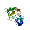
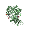
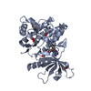
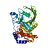
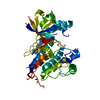
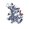
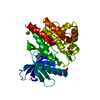
Method: torsion angle dynamics / Software ordinal: 1 Details: 15000 steps of refinement. Explicit distance restraints to the TEMPO spin-labeled reducing end of heparin dp8 were obtained from the PREs (T2) using an equation from Battiste and Wagner ...Details: 15000 steps of refinement. Explicit distance restraints to the TEMPO spin-labeled reducing end of heparin dp8 were obtained from the PREs (T2) using an equation from Battiste and Wagner (2000, Biochemistry) simplified by WHTC greater than1 to the form r equal to 4KTC/T2 given by Koppisetti et al. (2014, Nature Comms.). The rotational correlation time TC of the protein-heparin complexes was obtained from amide 15N cross-correlation rates nxy (Liu and Prestegard, 2008, J. Magn. Reson.) interpreted by the spectral density expression used in the TRACT approach (Lee et al., 2006. J. Magn. Reson.). Upper bounds were set at 15% above the distance estimate. An intermolecular NOE from an arginine backbone amide from 2H/15N-labeled enzyme to the anomeric proton H1 of GlcNS6S 5 or 7 had an upper bound of 6 A. Lower bounds were implicitly at van der Waals distance. Ambiguous distance restraints from any residue of heparin dp8 to explicit protein amide groups were applied on the basis of amide chemical shift perturbations from heparin dp8 or protection of amides by heparin dp4 from line broadening by Gd.EDTA with deltaT2 greater than 37/sec. Ambiguous distance restraints from any residue of heparin dp8 to lysine amino or arginine ureido groups were also applied on the basis of mutations that impaired activation of proMMP-7 by heparin dp16. The combination of the intermolecular distance restraints from the explicit PRE and NOE measurements and the ambiguous sources were used to dock coordinates of heparin dp8 with the NMR solution structure of human proMMP-7 of Prior et al. (2015, Structure). To enable molecular flexibility widely through the proMMP-7 and heparin dp8 chains while maintaining the structural integrity of the proMMP-7 during the restrained docking simulations, CYANA 2.1 (Guntert and Buchner, 2015, J. Biomol. NMR) was utilized in concert with the intramolecular proMMP-7 NOE-derived distance restraints and chemical shift-derived dihedral restraints of the NMR structure (Prior et al., 2015, Structure). CYANA topology files describing the sugar monomers were derived from topology files curated by the Automated Topology Builder (Malde et al., 2011, J. Chem. Theory Comput.) and based on accession number 9804 for 2-O-sulfo-alpha-L-idopyranuronic acid (IDS or IdoA2S) and 9778 for N,O6-disulfo-glucosamine (SGN or GlcNS6S). The globally flexible docking simulations required the proMMP-7 polypeptide to be linked to the calcium and zinc ions and heparin dp8 chains via tethers of non-interacting pseudoatoms. With this tethering and flexible structural integrity enforced, the carbohydrate chains were docked with the intermolecular distance restraints
NMR representative
Selection criteria: target function
NMR ensemble
Conformer selection criteria: structures with the least restraint violations Conformers calculated total number: 200 / Conformers submitted total number: 16
+
About Yorodumi
-
News
-
Feb 9, 2022. New format data for meta-information of EMDB entries
New format data for meta-information of EMDB entries
Version 3 of the EMDB header file is now the official format.
The previous official version 1.9 will be removed from the archive.
In the structure databanks used in Yorodumi, some data are registered as the other names, "COVID-19 virus" and "2019-nCoV". Here are the details of the virus and the list of structure data.
Jan 31, 2019. EMDB accession codes are about to change! (news from PDBe EMDB page)
EMDB accession codes are about to change! (news from PDBe EMDB page)
The allocation of 4 digits for EMDB accession codes will soon come to an end. Whilst these codes will remain in use, new EMDB accession codes will include an additional digit and will expand incrementally as the available range of codes is exhausted. The current 4-digit format prefixed with “EMD-” (i.e. EMD-XXXX) will advance to a 5-digit format (i.e. EMD-XXXXX), and so on. It is currently estimated that the 4-digit codes will be depleted around Spring 2019, at which point the 5-digit format will come into force.
The EM Navigator/Yorodumi systems omit the EMD- prefix.
Related info.:Q: What is EMD? / ID/Accession-code notation in Yorodumi/EM Navigator
Yorodumi is a browser for structure data from EMDB, PDB, SASBDB, etc.
This page is also the successor to EM Navigator detail page, and also detail information page/front-end page for Omokage search.
The word "yorodu" (or yorozu) is an old Japanese word meaning "ten thousand". "mi" (miru) is to see.
Related info.:EMDB / PDB / SASBDB / Comparison of 3 databanks / Yorodumi Search / Aug 31, 2016. New EM Navigator & Yorodumi / Yorodumi Papers / Jmol/JSmol / Function and homology information / Changes in new EM Navigator and Yorodumi
 Movie
Movie Controller

 Yorodumi
 Open data
 Basic information
 Components
 Keywords
 Function and homology information
 Homo sapiens (human)
 Authors
 United States, 1items
  Citation
 Structure visualization
 Downloads & links
 Other downloads


 PDBj
PDBj


 Assembly
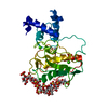


 Mass: 40.078 Da / Num. of mol.: 2 / Source method: obtained synthetically / Formula: Ca
 Mass: 65.409 Da / Num. of mol.: 2 / Source method: obtained synthetically / Formula: Zn
 Sample preparation
 Processing
 CYANA
CYANA