 Movie
Movie Controller

[English] 日本語
 Yorodumi
- PDB-5u9l: Crystal structure of the intermembrane space region of the plasti... -
+ Open data

- Basic information
| Entry | Database: PDB / ID: 5u9l | ||||||
|---|---|---|---|---|---|---|---|
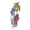
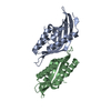
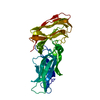
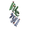
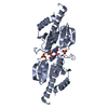
| Title | Crystal structure of the intermembrane space region of the plastid division protein PARC6 | ||||||
 Components Components | PARALOG OF ACCUMULATION AND REPLICATION OF CHLOROPLASTS 6 (PARC6) | ||||||
 Keywords Keywords | CELL CYCLE / Plastid / cell division / chloroplast / intermembrane space | ||||||
| Function / homology |  Function and homology information Function and homology informationplastid inner membrane / plastid fission / chloroplast fission / chloroplast inner membrane / chloroplast organization / chloroplast Similarity search - Function | ||||||
| Biological species |  | ||||||
| Method |  X-RAY DIFFRACTION / SYNCHROTRON / Resolution: 2.516 Å X-RAY DIFFRACTION / SYNCHROTRON / Resolution: 2.516 Å | ||||||
 Authors Authors | Delmar, J.D. / Chou, T.H. | ||||||
 Citation Citation | Journal: To Be Published Title: Cocrystal structure of the intermembrane space region of the plastid division proteins PARC6 and PDV1 Authors: Delmar, J.A. / Yu, E.W. / Osteryoung, K.W. | ||||||
| History |
|
- Structure visualization
| Structure viewer | Molecule: MolmilJmol/JSmol |
|---|
- Downloads & links
-Download
| PDBx/mmCIF format | 5u9l.cif.gz | 141.4 KB | Display | PDBx/mmCIF format |
|---|---|---|---|---|
| PDB format | pdb5u9l.ent.gz | 109.1 KB | Display | PDB format |
| PDBx/mmJSON format | 5u9l.json.gz | Tree view | PDBx/mmJSON format | |
| Others |  Other downloads Other downloads |
-Validation report
| Arichive directory | https://data.pdbj.org/pub/pdb/validation_reports/u9/5u9lftp://data.pdbj.org/pub/pdb/validation_reports/u9/5u9l | HTTPS FTP |
|---|
-Related structure data


-Links
 PDBj
PDBj- Assembly
| Deposited unit | 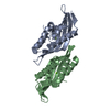
| |||||||||
|---|---|---|---|---|---|---|---|---|---|---|
| 1 |
| |||||||||
| Unit cell |
| |||||||||
| Components on special symmetry positions |
|
-Components
| #1: Protein | Mass: 26533.930 Da / Num. of mol.: 2 Source method: isolated from a genetically manipulated source Source: (gene. exp.)  #2: Chemical |   Mass: 24.305 Da / Num. of mol.: 2 / Source method: obtained synthetically / Formula: Mg Mass: 24.305 Da / Num. of mol.: 2 / Source method: obtained synthetically / Formula: Mg#3: Water | ChemComp-HOH / |  Mass: 18.015 Da / Num. of mol.: 53 / Source method: isolated from a natural source / Formula: H2O Mass: 18.015 Da / Num. of mol.: 53 / Source method: isolated from a natural source / Formula: H2OHas protein modification | Y | |
|---|
-Experimental details
-Experiment
| Experiment | Method: X-RAY DIFFRACTION / Number of used crystals: 1 |
|---|
- Sample preparation
| Crystal | Density Matthews: 1.7 Å3/Da / Density % sol: 27.6 % |
|---|---|
| Crystal grow | Temperature: 294 K / Method: vapor diffusion, hanging drop Details: 18% polyethylene glycol (PEG) 4000, 0.1 M Magnesium acetate, 0.1 M Na-MES (pH 6.5), 0.05 M Potassium nitrate and 5% glycerol |
-Data collection
| Diffraction | Mean temperature: 100 K | ||||||||||||||||||||||||||||||||||||||||||||||||||||||||||||
|---|---|---|---|---|---|---|---|---|---|---|---|---|---|---|---|---|---|---|---|---|---|---|---|---|---|---|---|---|---|---|---|---|---|---|---|---|---|---|---|---|---|---|---|---|---|---|---|---|---|---|---|---|---|---|---|---|---|---|---|---|---|
| Diffraction source | Source: SYNCHROTRON / Site: APS  / Beamline: 24-ID-C / Wavelength: 0.979 Å / Beamline: 24-ID-C / Wavelength: 0.979 Å | ||||||||||||||||||||||||||||||||||||||||||||||||||||||||||||
| Detector | Type: DECTRIS PILATUS 6M-F / Detector: PIXEL / Date: Feb 14, 2016 | ||||||||||||||||||||||||||||||||||||||||||||||||||||||||||||
| Radiation | Protocol: SINGLE WAVELENGTH / Monochromatic (M) / Laue (L): M / Scattering type: x-ray | ||||||||||||||||||||||||||||||||||||||||||||||||||||||||||||
| Radiation wavelength | Wavelength: 0.979 Å / Relative weight: 1 | ||||||||||||||||||||||||||||||||||||||||||||||||||||||||||||
| Reflection | Resolution: 2.5→40 Å / Num. obs: 12543 / % possible obs: 99.5 % / Redundancy: 8.3 % / Rmerge(I) obs: 0.139 / Net I/σ(I): 6.2 | ||||||||||||||||||||||||||||||||||||||||||||||||||||||||||||
| Reflection shell |
|
- Processing
| Software |
| ||||||||||||||||||||||||||||||||||||||||
|---|---|---|---|---|---|---|---|---|---|---|---|---|---|---|---|---|---|---|---|---|---|---|---|---|---|---|---|---|---|---|---|---|---|---|---|---|---|---|---|---|---|
| Refinement | Resolution: 2.516→39.91 Å / SU ML: 0.29 / Cross valid method: FREE R-VALUE / σ(F): 1.35 / Phase error: 28
| ||||||||||||||||||||||||||||||||||||||||
| Solvent computation | Shrinkage radii: 0.9 Å / VDW probe radii: 1.11 Å | ||||||||||||||||||||||||||||||||||||||||
| Displacement parameters | Biso max: 165.26 Å2 / Biso mean: 65.9137 Å2 / Biso min: 27.85 Å2 | ||||||||||||||||||||||||||||||||||||||||
| Refinement step | Cycle: final / Resolution: 2.516→39.91 Å
| ||||||||||||||||||||||||||||||||||||||||
| Refinement TLS params. | Method: refined / Origin x: -8.7325 Å / Origin y: -17.6226 Å / Origin z: -0.9824 Å
| ||||||||||||||||||||||||||||||||||||||||
| Refinement TLS group |
|
