 Movie
Movie Controller

[English] 日本語
 Yorodumi
- PDB-5u7v: Crystal structure of a nucleoside triphosphate diphosphohydrolase... -
+ Open data

- Basic information
| Entry | Database: PDB / ID: 5u7v | ||||||
|---|---|---|---|---|---|---|---|
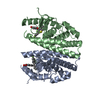
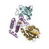
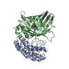
| Title | Crystal structure of a nucleoside triphosphate diphosphohydrolase (NTPDase) from the legume Trifolium repens in complex with AMP | ||||||
 Components Components | Apyrase | ||||||
 Keywords Keywords | HYDROLASE / apyrase / NTPDase / RNase-H fold / mixed 5 strand beta-sheet | ||||||
| Function / homology |  Function and homology information Function and homology informationapyrase / apyrase activity / nucleoside diphosphate catabolic process / nucleoside diphosphate phosphatase activity / ATP binding / membrane Similarity search - Function | ||||||
| Biological species |  Trifolium repens (white clover) Trifolium repens (white clover) | ||||||
| Method |  X-RAY DIFFRACTION / SYNCHROTRON / MOLECULAR REPLACEMENT / Resolution: 2.15 Å X-RAY DIFFRACTION / SYNCHROTRON / MOLECULAR REPLACEMENT / Resolution: 2.15 Å | ||||||
 Authors Authors | Cumming, M.H. / Summers, E.L. / Oulavallickal, T. / Roberts, N. / Arcus, V.L. | ||||||
 Citation Citation | Journal: Protein Sci. / Year: 2017 Title: Structures and kinetics for plant nucleoside triphosphate diphosphohydrolases support a domain motion catalytic mechanism. Authors: Summers, E.L. / Cumming, M.H. / Oulavallickal, T. / Roberts, N.J. / Arcus, V.L. | ||||||
| History |
|
- Structure visualization
| Structure viewer | Molecule: MolmilJmol/JSmol |
|---|
- Downloads & links
-Download
| PDBx/mmCIF format | 5u7v.cif.gz | 132.7 KB | Display | PDBx/mmCIF format |
|---|---|---|---|---|
| PDB format | pdb5u7v.ent.gz | 98.2 KB | Display | PDB format |
| PDBx/mmJSON format | 5u7v.json.gz | Tree view | PDBx/mmJSON format | |
| Others |  Other downloads Other downloads |
-Validation report
| Summary document | 5u7v_validation.pdf.gz | 718.5 KB | Display | wwPDB validaton report |
|---|---|---|---|---|
| Full document | 5u7v_full_validation.pdf.gz | 723.4 KB | Display | |
| Data in XML | 5u7v_validation.xml.gz | 18 KB | Display | |
| Data in CIF | 5u7v_validation.cif.gz | 26 KB | Display | |
| Arichive directory | https://data.pdbj.org/pub/pdb/validation_reports/u7/5u7vftp://data.pdbj.org/pub/pdb/validation_reports/u7/5u7v | HTTPS FTP |
-Related structure data
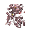
| Related structure data | 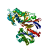 5u7pSC 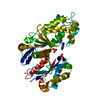 5u7wC  5u7xC C: citing same article ( S: Starting model for refinement |
|---|---|
| Similar structure data |


-Links
 PDBj
PDBj


- Assembly
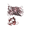
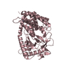
| Deposited unit | 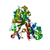
| ||||||||
|---|---|---|---|---|---|---|---|---|---|
| 1 |
| ||||||||
| Unit cell |
|
-Components
| #1: Protein | Mass: 46992.301 Da / Num. of mol.: 1 / Fragment: residues 39-455 Source method: isolated from a genetically manipulated source Source: (gene. exp.) Trifolium repens (white clover) / Production host:  |
|---|---|
| #2: Chemical | ChemComp-AMP /   Mass: 347.221 Da / Num. of mol.: 1 / Source method: obtained synthetically / Formula: C10H14N5O7P / Comment: AMP*YM Mass: 347.221 Da / Num. of mol.: 1 / Source method: obtained synthetically / Formula: C10H14N5O7P / Comment: AMP*YM |
| #3: Water | ChemComp-HOH /  Mass: 18.015 Da / Num. of mol.: 158 / Source method: isolated from a natural source / Formula: H2O Mass: 18.015 Da / Num. of mol.: 158 / Source method: isolated from a natural source / Formula: H2O |
| Has protein modification | Y |
-Experimental details
-Experiment
| Experiment | Method: X-RAY DIFFRACTION / Number of used crystals: 1 |
|---|
- Sample preparation
| Crystal | Density Matthews: 1.96 Å3/Da / Density % sol: 37.21 % / Description: large thick plates |
|---|---|
| Crystal grow | Temperature: 291 K / Method: vapor diffusion, hanging drop / pH: 8 Details: 0.2 M ammonium dihydrogen orthophosphate, 15% PEG 3350 |
-Data collection
| Diffraction | Mean temperature: 100 K |
|---|---|
| Diffraction source | Source: SYNCHROTRON / Site: Australian Synchrotron  / Beamline: MX2 / Wavelength: 0.9537 Å / Beamline: MX2 / Wavelength: 0.9537 Å |
| Detector | Type: ADSC QUANTUM 315r / Detector: CCD / Date: Oct 1, 2009 |
| Radiation | Protocol: SINGLE WAVELENGTH / Monochromatic (M) / Laue (L): M / Scattering type: x-ray |
| Radiation wavelength | Wavelength: 0.9537 Å / Relative weight: 1 |
| Reflection | Resolution: 2.15→65.9 Å / Num. obs: 19997 / % possible obs: 100 % / Redundancy: 3.6 % / Biso Wilson estimate: 32.43 Å2 / Rmerge(I) obs: 0.136 / Net I/σ(I): 6.2 |
| Reflection shell | Redundancy: 3.7 % / Rmerge(I) obs: 0.462 / Mean I/σ(I) obs: 2.5 / % possible all: 100 |
- Processing
| Software |
| ||||||||||||||||||||||||||||||||||||||||||||||||||||||||||||||||||||||||||||||||||||||||||||||||||||||||||||||||||||||||||||||||||||||||||||||||||||||||||||||||||||||||||||||||||||||
|---|---|---|---|---|---|---|---|---|---|---|---|---|---|---|---|---|---|---|---|---|---|---|---|---|---|---|---|---|---|---|---|---|---|---|---|---|---|---|---|---|---|---|---|---|---|---|---|---|---|---|---|---|---|---|---|---|---|---|---|---|---|---|---|---|---|---|---|---|---|---|---|---|---|---|---|---|---|---|---|---|---|---|---|---|---|---|---|---|---|---|---|---|---|---|---|---|---|---|---|---|---|---|---|---|---|---|---|---|---|---|---|---|---|---|---|---|---|---|---|---|---|---|---|---|---|---|---|---|---|---|---|---|---|---|---|---|---|---|---|---|---|---|---|---|---|---|---|---|---|---|---|---|---|---|---|---|---|---|---|---|---|---|---|---|---|---|---|---|---|---|---|---|---|---|---|---|---|---|---|---|---|---|---|
| Refinement | Method to determine structure: MOLECULAR REPLACEMENT Starting model: 5u7p Resolution: 2.15→35.43 Å / Cor.coef. Fo:Fc: 0.947 / Cor.coef. Fo:Fc free: 0.912 / SU B: 13.357 / SU ML: 0.176 / Cross valid method: THROUGHOUT / ESU R: 0.304 / ESU R Free: 0.237 / Stereochemistry target values: MAXIMUM LIKELIHOOD / Details: HYDROGENS HAVE BEEN ADDED IN THE RIDING POSITIONS
| ||||||||||||||||||||||||||||||||||||||||||||||||||||||||||||||||||||||||||||||||||||||||||||||||||||||||||||||||||||||||||||||||||||||||||||||||||||||||||||||||||||||||||||||||||||||
| Solvent computation | Ion probe radii: 0.8 Å / Shrinkage radii: 0.8 Å / VDW probe radii: 1.2 Å / Solvent model: MASK | ||||||||||||||||||||||||||||||||||||||||||||||||||||||||||||||||||||||||||||||||||||||||||||||||||||||||||||||||||||||||||||||||||||||||||||||||||||||||||||||||||||||||||||||||||||||
| Displacement parameters | Biso mean: 34.075 Å2
| ||||||||||||||||||||||||||||||||||||||||||||||||||||||||||||||||||||||||||||||||||||||||||||||||||||||||||||||||||||||||||||||||||||||||||||||||||||||||||||||||||||||||||||||||||||||
| Refinement step | Cycle: 1 / Resolution: 2.15→35.43 Å
| ||||||||||||||||||||||||||||||||||||||||||||||||||||||||||||||||||||||||||||||||||||||||||||||||||||||||||||||||||||||||||||||||||||||||||||||||||||||||||||||||||||||||||||||||||||||
| Refine LS restraints |
|