[English] 日本語
Yorodumi
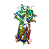
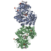
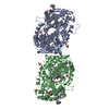
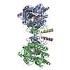
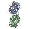
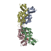
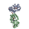
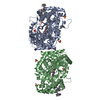
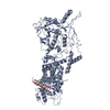
- PDB-5u73: Crystal structure of human Niemann-Pick C1 protein -

+
Open data


ID or keywords:

Loading...

-
Basic information

Entry
Database: PDB / ID: 5u73
TitleCrystal structure of human Niemann-Pick C1 protein
ComponentsNiemann-Pick C1 protein
KeywordsMEMBRANE PROTEIN
Function / homology
Function and homology information


cyclodextrin metabolic process / cholesterol storage / membrane raft organization / : / intracellular cholesterol transport / intracellular lipid transport / intestinal cholesterol absorption / LDL clearance / programmed cell death / negative regulation of epithelial cell apoptotic process ...cyclodextrin metabolic process / cholesterol storage / membrane raft organization / : / intracellular cholesterol transport / intracellular lipid transport / intestinal cholesterol absorption / LDL clearance / programmed cell death / negative regulation of epithelial cell apoptotic process / cholesterol transport / bile acid metabolic process / establishment of protein localization to membrane / cholesterol efflux / adult walking behavior / lysosomal transport / cholesterol binding / negative regulation of macroautophagy / cellular response to steroid hormone stimulus / protein glycosylation / cellular response to low-density lipoprotein particle stimulus / response to cadmium ion / negative regulation of TORC1 signaling / cholesterol metabolic process / neurogenesis / cholesterol homeostasis / liver development / macroautophagy / autophagy / endocytosis / transmembrane signaling receptor activity / virus receptor activity / signaling receptor activity / nuclear envelope / late endosome membrane / gene expression / lysosome / response to xenobiotic stimulus / symbiont entry into host cell / membrane raft / lysosomal membrane / perinuclear region of cytoplasm / Golgi apparatus / endoplasmic reticulum / extracellular exosome / extracellular region / membrane / plasma membrane
Similarity search - Function
NPC1-like / Niemann-Pick C1, N-terminal / Niemann-Pick C1 N terminus / Sterol-sensing domain of SREBP cleavage-activation / Protein patched/dispatched / Patched family / Sterol-sensing domain (SSD) profile. / Sterol-sensing domain
Similarity search - Domain/homology
NPC intracellular cholesterol transporter 1
Similarity search - Component
Biological speciesHomo sapiens (human)
MethodX-RAY DIFFRACTION / SYNCHROTRON / SAD / Resolution: 3.348 Å
AuthorsLi, X. / Wang, J. / Blobel, G.
CitationJournal: Proc. Natl. Acad. Sci. U.S.A. / Year: 2017
Title: 3.3 angstrom structure of Niemann-Pick C1 protein reveals insights into the function of the C-terminal luminal domain in cholesterol transport.
Authors: Li, X. / Lu, F. / Trinh, M.N. / Schmiege, P. / Seemann, J. / Wang, J. / Blobel, G.
History
DepositionDec 11, 2016Deposition site: RCSB / Processing site: RCSB
Revision 1.0Sep 27, 2017Provider: repository / Type: Initial release
SupersessionOct 11, 2017ID: 5I31
Revision 1.1Oct 11, 2017Group: Advisory / Structure summary / Category: entity / pdbx_database_PDB_obs_spr / Item: _entity.src_method
Revision 2.0Jul 29, 2020Group: Advisory / Atomic model ...Advisory / Atomic model / Data collection / Derived calculations / Structure summary
Category: atom_site / chem_comp ...atom_site / chem_comp / entity / pdbx_branch_scheme / pdbx_chem_comp_identifier / pdbx_entity_branch / pdbx_entity_branch_descriptor / pdbx_entity_branch_link / pdbx_entity_branch_list / pdbx_entity_nonpoly / pdbx_nonpoly_scheme / pdbx_struct_assembly_gen / pdbx_validate_close_contact / struct_asym / struct_conn / struct_site / struct_site_gen
Item: _atom_site.B_iso_or_equiv / _atom_site.Cartn_x ..._atom_site.B_iso_or_equiv / _atom_site.Cartn_x / _atom_site.Cartn_y / _atom_site.Cartn_z / _atom_site.auth_asym_id / _atom_site.auth_atom_id / _atom_site.auth_comp_id / _atom_site.auth_seq_id / _atom_site.label_asym_id / _atom_site.label_atom_id / _atom_site.label_comp_id / _atom_site.label_entity_id / _atom_site.type_symbol / _chem_comp.name / _chem_comp.type / _pdbx_struct_assembly_gen.asym_id_list / _pdbx_validate_close_contact.auth_asym_id_1 / _pdbx_validate_close_contact.auth_asym_id_2 / _pdbx_validate_close_contact.auth_seq_id_1 / _pdbx_validate_close_contact.auth_seq_id_2 / _struct_conn.pdbx_role / _struct_conn.ptnr1_auth_asym_id / _struct_conn.ptnr1_auth_seq_id / _struct_conn.ptnr1_label_asym_id / _struct_conn.ptnr2_auth_asym_id / _struct_conn.ptnr2_auth_seq_id / _struct_conn.ptnr2_label_asym_id
Description: Carbohydrate remediation / Provider: repository / Type: Remediation

-
Structure visualization

Structure viewerMolecule:
MolmilJmol/JSmol

Downloads & links

-
Assembly

Deposited unit
A: Niemann-Pick C1 protein
hetero molecules


Theoretical massNumber of molelcules
Total (without water)145,62212
Polymers142,2551
Non-polymers3,36711
Water0
1


  • Idetical with deposited unit
  • defined by author&software
TypeNameSymmetry operationNumber
identity operation1_555x,y,z1
MethodPISA
Unit cell
Length a, b, c (Å)181.937, 223.071, 63.208
Angle α, β, γ (deg.)90.00, 105.31, 90.00
Int Tables number5
Space group name H-MC121

-
Components

#1: Protein Niemann-Pick C1 protein


Mass: 142254.859 Da / Num. of mol.: 1
Source method: isolated from a genetically manipulated source
Source: (gene. exp.) Homo sapiens (human) / Gene: NPC1 / Production host: Homo sapiens (human) / References: UniProt: O15118
#2: Polysaccharide 2-acetamido-2-deoxy-beta-D-glucopyranose-(1-4)-2-acetamido-2-deoxy-beta-D-glucopyranose


Type: oligosaccharide / Mass: 424.401 Da / Num. of mol.: 1 / Source method: obtained synthetically
DescriptorTypeProgram
DGlcpNAcb1-4DGlcpNAcb1-Glycam Condensed SequenceGMML 1.0
WURCS=2.0/1,2,1/[a2122h-1b_1-5_2*NCC/3=O]/1-1/a4-b1WURCSPDB2Glycan 1.1.0
[]{[(4+1)][b-D-GlcpNAc]{[(4+1)][b-D-GlcpNAc]{}}}LINUCSPDB-CARE
#3: Polysaccharide alpha-D-mannopyranose-(1-4)-2-acetamido-2-deoxy-beta-D-glucopyranose-(1-4)-2-acetamido-2-deoxy-beta- ...alpha-D-mannopyranose-(1-4)-2-acetamido-2-deoxy-beta-D-glucopyranose-(1-4)-2-acetamido-2-deoxy-beta-D-glucopyranose


Type: oligosaccharide / Mass: 586.542 Da / Num. of mol.: 2 / Source method: obtained synthetically
DescriptorTypeProgram
DManpa1-4DGlcpNAcb1-4DGlcpNAcb1-Glycam Condensed SequenceGMML 1.0
WURCS=2.0/2,3,2/[a2122h-1b_1-5_2*NCC/3=O][a1122h-1a_1-5]/1-1-2/a4-b1_b4-c1WURCSPDB2Glycan 1.1.0
[]{[(4+1)][b-D-GlcpNAc]{[(4+1)][b-D-GlcpNAc]{[(4+1)][a-D-Manp]{}}}}LINUCSPDB-CARE
#4: Sugar
ChemComp-NAG / 2-acetamido-2-deoxy-beta-D-glucopyranose / N-Acetylglucosamine


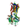
Type: D-saccharide, beta linking / Mass: 221.208 Da / Num. of mol.: 8 / Source method: obtained synthetically / Formula: C8H15NO6
IdentifierTypeProgram
DGlcpNAcbCONDENSED IUPAC CARBOHYDRATE SYMBOLGMML 1.0
N-acetyl-b-D-glucopyranosamineCOMMON NAMEGMML 1.0
b-D-GlcpNAcIUPAC CARBOHYDRATE SYMBOLPDB-CARE 1.0
GlcNAcSNFG CARBOHYDRATE SYMBOLGMML 1.0

-
Experimental details

-
Experiment

ExperimentMethod: X-RAY DIFFRACTION / Number of used crystals: 1

-
Sample preparation

CrystalDensity Matthews: 4.35 Å3/Da / Density % sol: 71.71 %
Crystal growTemperature: 293 K / Method: evaporation / pH: 5.5
Details: 0.1 M sodium citrate pH 5.5 , 0.1 M NaCl, 12% (vol/vol) PEG4000

-
Data collection

DiffractionMean temperature: 100 K
Diffraction sourceSource: SYNCHROTRON / Site: ALS / Beamline: 8.2.1 / Wavelength: 1 Å
DetectorType: MARMOSAIC 325 mm CCD / Detector: CCD / Date: Aug 30, 2015
RadiationProtocol: SINGLE WAVELENGTH / Monochromatic (M) / Laue (L): M / Scattering type: x-ray
Radiation wavelengthWavelength: 1 Å / Relative weight: 1
ReflectionResolution: 3.348→50 Å / Num. obs: 17949 / % possible obs: 51.5 % / Redundancy: 3 % / Net I/σ(I): 9.5

-
Processing

Software
NameVersionClassification
PHENIX(1.11.1_2575: ???)refinement
HKL-2000data scaling
HKL-2000data reduction
PHASERphasing
RefinementMethod to determine structure: SAD / Resolution: 3.348→41.148 Å / SU ML: 0.51 / Cross valid method: FREE R-VALUE / σ(F): 1.37 / Phase error: 30.43 / Stereochemistry target values: ML
RfactorNum. reflection% reflection
Rfree0.2576 873 4.86 %
Rwork0.2327 --
obs0.234 17949 51.48 %
Solvent computationShrinkage radii: 0.9 Å / VDW probe radii: 1.11 Å / Solvent model: FLAT BULK SOLVENT MODEL
Refinement stepCycle: LAST / Resolution: 3.348→41.148 Å
ProteinNucleic acidLigandSolventTotal
Num. atoms7044 0 218 0 7262
Refine LS restraints
Refine-IDTypeDev idealNumber
X-RAY DIFFRACTIONf_bond_d0.0047462
X-RAY DIFFRACTIONf_angle_d2.03410198
X-RAY DIFFRACTIONf_dihedral_angle_d15.0344344
X-RAY DIFFRACTIONf_chiral_restr0.0671210
X-RAY DIFFRACTIONf_plane_restr0.0041272
LS refinement shell
Resolution (Å)Rfactor RfreeNum. reflection RfreeRfactor RworkNum. reflection RworkRefine-ID% reflection obs (%)
3.3484-3.55810.564660.2732232X-RAY DIFFRACTION4
3.5581-3.83260.2658340.235841X-RAY DIFFRACTION15
3.8326-4.2180.24581240.21641999X-RAY DIFFRACTION36
4.218-4.82750.23261610.19213442X-RAY DIFFRACTION62
4.8275-6.0790.26372490.24195034X-RAY DIFFRACTION91
6.079-41.15150.26662990.25785528X-RAY DIFFRACTION99
Refinement TLS params.

Method: refined / Refine-ID: X-RAY DIFFRACTION

IDL112)L122)L132)L222)L232)L332)S11 (Å °)S12 (Å °)S13 (Å °)S21 (Å °)S22 (Å °)S23 (Å °)S31 (Å °)S32 (Å °)S33 (Å °)T112)T122)T132)T222)T232)T332)Origin x (Å)Origin y (Å)Origin z (Å)
10.59660.4781-0.12041.24820.22180.74890.1569-0.19340.07880.3177-0.24080.591-0.0289-0.2228-0.00580.37760.02310.14650.30690.00030.322741.632274.354141.4551
22.11780.5972-0.06711.868-0.7692.8431-0.0154-0.2212-0.3297-0.0377-0.21380.1820.1662-0.0020.12460.239-0.02150.18860.1754-0.02750.371146.522888.701336.5322
30.75150.1045-0.28551.415-0.68950.8882-0.2430.2265-0.0177-0.20530.13080.77440.5845-0.38410.07990.6447-0.23070.15170.415-0.12270.577628.90736.927236.7839
41.9865-0.0150.01331.82380.18041.5-0.1215-0.2117-0.3709-0.2475-0.04730.3668-0.0393-0.1977-0.62830.16630.0114-0.00010.07810.03120.057652.572874.727532.9815
50.8101-0.0291-0.80180.9651-0.4830.9874-0.206-0.0125-0.1874-0.24390.14950.1850.174-0.01380.02990.20930.0356-0.09120.19890.01660.075353.172160.461432.2384
Refinement TLS group
IDRefine-IDRefine TLS-IDSelection details
1X-RAY DIFFRACTION1chain 'A' and (resid 334 through 508 )
2X-RAY DIFFRACTION2chain 'A' and (resid 509 through 592 )
3X-RAY DIFFRACTION3chain 'A' and (resid 593 through 861 )
4X-RAY DIFFRACTION4chain 'A' and (resid 862 through 908 )
5X-RAY DIFFRACTION5chain 'A' and (resid 909 through 1254 )

+
About Yorodumi

-
News

-
Feb 9, 2022. New format data for meta-information of EMDB entries

New format data for meta-information of EMDB entries

  • Version 3 of the EMDB header file is now the official format.
  • The previous official version 1.9 will be removed from the archive.

Related info.:EMDB header

External links:wwPDB to switch to version 3 of the EMDB data model

-
Aug 12, 2020. Covid-19 info

Covid-19 info

URL: https://pdbj.org/emnavi/covid19.php

New page: Covid-19 featured information page in EM Navigator.

Related info.:Covid-19 info / Mar 5, 2020. Novel coronavirus structure data

+
Mar 5, 2020. Novel coronavirus structure data

Novel coronavirus structure data

Related info.:Yorodumi Speices / Aug 12, 2020. Covid-19 info

External links:COVID-19 featured content - PDBj / Molecule of the Month (242):Coronavirus Proteases

+
Jan 31, 2019. EMDB accession codes are about to change! (news from PDBe EMDB page)

EMDB accession codes are about to change! (news from PDBe EMDB page)

  • The allocation of 4 digits for EMDB accession codes will soon come to an end. Whilst these codes will remain in use, new EMDB accession codes will include an additional digit and will expand incrementally as the available range of codes is exhausted. The current 4-digit format prefixed with “EMD-” (i.e. EMD-XXXX) will advance to a 5-digit format (i.e. EMD-XXXXX), and so on. It is currently estimated that the 4-digit codes will be depleted around Spring 2019, at which point the 5-digit format will come into force.
  • The EM Navigator/Yorodumi systems omit the EMD- prefix.

Related info.:Q: What is EMD? / ID/Accession-code notation in Yorodumi/EM Navigator

External links:EMDB Accession Codes are Changing Soon! / Contact to PDBj

+
Jul 12, 2017. Major update of PDB

Major update of PDB

  • wwPDB released updated PDB data conforming to the new PDBx/mmCIF dictionary.
  • This is a major update changing the version number from 4 to 5, and with Remediation, in which all the entries are updated.
  • In this update, many items about electron microscopy experimental information are reorganized (e.g. em_software).
  • Now, EM Navigator and Yorodumi are based on the updated data.

External links:wwPDB Remediation / Enriched Model Files Conforming to OneDep Data Standards Now Available in the PDB FTP Archive

-
Yorodumi

Thousand views of thousand structures

  • Yorodumi is a browser for structure data from EMDB, PDB, SASBDB, etc.
  • This page is also the successor to EM Navigator detail page, and also detail information page/front-end page for Omokage search.
  • The word "yorodu" (or yorozu) is an old Japanese word meaning "ten thousand". "mi" (miru) is to see.

Related info.:EMDB / PDB / SASBDB / Comparison of 3 databanks / Yorodumi Search / Aug 31, 2016. New EM Navigator & Yorodumi / Yorodumi Papers / Jmol/JSmol / Function and homology information / Changes in new EM Navigator and Yorodumi

Read more