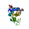
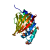
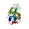
[English] 日本語
Yorodumi
- PDB-5u1m: Structure of the IRS-1 PTB Domain Bound to the Juxtamembrane Regi... -

+
Open data


ID or keywords:

Loading...

-
Basic information

Entry
Database: PDB / ID: 5u1m
TitleStructure of the IRS-1 PTB Domain Bound to the Juxtamembrane Region of the Insulin Receptor
Components
  • Insulin receptor
  • Insulin receptor substrate 1
KeywordsPROTEIN BINDING / PTB domain / Insulin Receptor Substrate-1 / Phosphopeptide / Insulin Receptor
Function / homology
Function and homology information


IRS-related events triggered by IGF1R / regulation of female gonad development / positive regulation of meiotic cell cycle / IRS-mediated signalling / positive regulation of fatty acid beta-oxidation / insulin-like growth factor II binding / positive regulation of developmental growth / male sex determination / insulin receptor complex / positive regulation of glucose metabolic process ...IRS-related events triggered by IGF1R / regulation of female gonad development / positive regulation of meiotic cell cycle / IRS-mediated signalling / positive regulation of fatty acid beta-oxidation / insulin-like growth factor II binding / positive regulation of developmental growth / male sex determination / insulin receptor complex / positive regulation of glucose metabolic process / insulin-like growth factor I binding / Activated NTRK3 signals through PI3K / transmembrane receptor protein tyrosine kinase adaptor activity / exocrine pancreas development / positive regulation of protein-containing complex disassembly / Signaling by Leptin / Signaling by LTK / dendritic spine maintenance / PI3K/AKT activation / cargo receptor activity / insulin binding / cellular response to fatty acid / neuronal cell body membrane / Signaling by ALK / adrenal gland development / PTB domain binding / Signaling by Insulin receptor / IRS activation / positive regulation of respiratory burst / amyloid-beta clearance / regulation of embryonic development / positive regulation of receptor internalization / protein kinase activator activity / insulin receptor substrate binding / PI3K Cascade / SOS-mediated signalling / epidermis development / positive regulation of glycogen biosynthetic process / Signal attenuation / phosphatidylinositol 3-kinase binding / positive regulation of insulin receptor signaling pathway / transport across blood-brain barrier / negative regulation of insulin secretion / Growth hormone receptor signaling / heart morphogenesis / activation of protein kinase B activity / Insulin receptor recycling / insulin-like growth factor receptor binding / signaling adaptor activity / dendrite membrane / phosphotyrosine residue binding / neuron projection maintenance / positive regulation of MAP kinase activity / Interleukin-7 signaling / negative regulation of insulin receptor signaling pathway / positive regulation of mitotic nuclear division / Insulin receptor signalling cascade / receptor-mediated endocytosis / SH2 domain binding / insulin-like growth factor receptor signaling pathway / positive regulation of glycolytic process / protein kinase C binding / learning / positive regulation of D-glucose import / phosphatidylinositol 3-kinase/protein kinase B signal transduction / response to insulin / insulin receptor binding / placental growth factor receptor activity / insulin receptor activity / vascular endothelial growth factor receptor activity / hepatocyte growth factor receptor activity / macrophage colony-stimulating factor receptor activity / platelet-derived growth factor alpha-receptor activity / platelet-derived growth factor beta-receptor activity / stem cell factor receptor activity / boss receptor activity / protein tyrosine kinase collagen receptor activity / brain-derived neurotrophic factor receptor activity / transmembrane-ephrin receptor activity / GPI-linked ephrin receptor activity / epidermal growth factor receptor activity / fibroblast growth factor receptor activity / cytokine-mediated signaling pathway / insulin-like growth factor receptor activity / receptor protein-tyrosine kinase / peptidyl-tyrosine phosphorylation / caveola / receptor internalization / memory / cellular response to insulin stimulus / male gonad development / Constitutive Signaling by Aberrant PI3K in Cancer / positive regulation of nitric oxide biosynthetic process / Signaling by ALK fusions and activated point mutants / late endosome / insulin receptor signaling pathway / glucose homeostasis / PIP3 activates AKT signaling / signaling receptor complex adaptor activity / amyloid-beta binding
Similarity search - Function
Insulin receptor substrate / Phosphotyrosine-binding domain (IRS1-like) / IRS-type PTB domain profile. / Phosphotyrosine-binding domain / IRS-type PTB domain / PTB domain (IRS-1 type) / Insulin receptor, trans-membrane domain / Insulin receptor trans-membrane segment / Tyrosine-protein kinase, insulin-like receptor / Tyrosine-protein kinase, receptor class II, conserved site ...Insulin receptor substrate / Phosphotyrosine-binding domain (IRS1-like) / IRS-type PTB domain profile. / Phosphotyrosine-binding domain / IRS-type PTB domain / PTB domain (IRS-1 type) / Insulin receptor, trans-membrane domain / Insulin receptor trans-membrane segment / Tyrosine-protein kinase, insulin-like receptor / Tyrosine-protein kinase, receptor class II, conserved site / Receptor tyrosine kinase class II signature. / Pleckstrin-homology domain (PH domain)/Phosphotyrosine-binding domain (PTB) / PH-domain like / Receptor L-domain / Furin-like cysteine-rich domain / Receptor L-domain superfamily / Furin-like cysteine rich region / Receptor L domain / Furin-like repeat / Furin-like repeats / PH domain / PH domain profile. / Pleckstrin homology domain. / Growth factor receptor cysteine-rich domain superfamily / Pleckstrin homology domain / Fibronectin type III domain / : / Fibronectin type 3 domain / Fibronectin type-III domain profile. / Fibronectin type III / Fibronectin type III superfamily / Tyrosine-protein kinase, catalytic domain / Tyrosine kinase, catalytic domain / Tyrosine protein kinases specific active-site signature. / Tyrosine-protein kinase, active site / PH-like domain superfamily / Serine-threonine/tyrosine-protein kinase, catalytic domain / Protein tyrosine and serine/threonine kinase / Roll / Protein kinase, ATP binding site / Protein kinases ATP-binding region signature. / Immunoglobulin-like fold / Protein kinase domain profile. / Protein kinase domain / Protein kinase-like domain superfamily / Mainly Beta
Similarity search - Domain/homology
Insulin receptor / Insulin receptor substrate 1
Similarity search - Component
Biological speciesHomo sapiens (human)
MethodX-RAY DIFFRACTION / Resolution: 1.8 Å
AuthorsEck, M.J. / Dhe-Paganon, S.
CitationJournal: Nat Commun / Year: 2017
Title: Domain-dependent effects of insulin and IGF-1 receptors on signalling and gene expression.
Authors: Cai, W. / Sakaguchi, M. / Kleinridders, A. / Gonzalez-Del Pino, G. / Dreyfuss, J.M. / O'Neill, B.T. / Ramirez, A.K. / Pan, H. / Winnay, J.N. / Boucher, J. / Eck, M.J. / Kahn, C.R.
History
DepositionNov 28, 2016Deposition site: RCSB / Processing site: RCSB
Revision 1.0Sep 13, 2017Provider: repository / Type: Initial release
Revision 1.1Nov 6, 2024Group: Data collection / Database references / Structure summary
Category: chem_comp_atom / chem_comp_bond ...chem_comp_atom / chem_comp_bond / database_2 / pdbx_entry_details / pdbx_modification_feature
Item: _database_2.pdbx_DOI / _database_2.pdbx_database_accession

-
Structure visualization

Structure viewerMolecule:
MolmilJmol/JSmol

Downloads & links

-
Assembly

Deposited unit
A: Insulin receptor substrate 1
B: Insulin receptor


Theoretical massNumber of molelcules
Total (without water)12,9242
Polymers12,9242
Non-polymers00
Water2,162120
1


  • Idetical with deposited unit
  • defined by author&software
TypeNameSymmetry operationNumber
identity operation1_555x,y,z1
Buried area1360 Å2
ΔGint-11 kcal/mol
Surface area6480 Å2
MethodPISA
Unit cell
Length a, b, c (Å)68.370, 68.370, 57.520
Angle α, β, γ (deg.)90.000, 90.000, 90.000
Int Tables number89
Space group name H-MP422

-
Components

#1: Protein Insulin receptor substrate 1 / IRS-1


Mass: 11832.777 Da / Num. of mol.: 1 / Fragment: PTB domain (UNP residues 161-265)
Source method: isolated from a genetically manipulated source
Source: (gene. exp.) Homo sapiens (human) / Gene: IRS1 / Production host: Escherichia coli K-12 (bacteria) / References: UniProt: P35568
#2: Protein/peptide Insulin receptor / IR


Mass: 1091.066 Da / Num. of mol.: 1 / Fragment: juxtamembrane region (UNP residues 991-999)
Source method: isolated from a genetically manipulated source
Source: (gene. exp.) Homo sapiens (human) / Gene: INSR / Production host: Escherichia coli K-12 (bacteria) / References: UniProt: P06213
#3: Water ChemComp-HOH / water


Mass: 18.015 Da / Num. of mol.: 120 / Source method: isolated from a natural source / Formula: H2O
Has protein modificationY

-
Experimental details

-
Experiment

ExperimentMethod: X-RAY DIFFRACTION / Number of used crystals: 1

-
Sample preparation

CrystalDensity Matthews: 2.6 Å3/Da / Density % sol: 52.7 %
Crystal growTemperature: 293 K / Method: vapor diffusion, hanging drop / Details: ammonium sulfate

-
Data collection

DiffractionMean temperature: 293 K
Diffraction sourceSource: ROTATING ANODE / Type: ELLIOTT GX-13 / Wavelength: 1 Å
DetectorType: MARRESEARCH / Detector: IMAGE PLATE / Date: Jan 1, 1996
RadiationProtocol: SINGLE WAVELENGTH / Monochromatic (M) / Laue (L): M / Scattering type: x-ray
Radiation wavelengthWavelength: 1 Å / Relative weight: 1
ReflectionResolution: 1.8→20 Å / Num. obs: 12453 / % possible obs: 94.9 % / Redundancy: 6 % / Net I/σ(I): 15
Reflection shellHighest resolution: 1.8 Å

-
Processing

Software
NameVersionClassification
REFMACrefinement
PDB_EXTRACT3.2data extraction
XDSdata reduction
XSCALEdata scaling
MLPHAREphasing
RefinementResolution: 1.8→20 Å / Cross valid method: FREE R-VALUE /
Num. reflection% reflection
obs12453 94.9 %
Displacement parametersBiso max: 51.58 Å2 / Biso mean: 12.4704 Å2 / Biso min: 0 Å2
Refinement stepCycle: LAST / Resolution: 1.8→20 Å
ProteinNucleic acidLigandSolventTotal
Num. atoms905 0 0 120 1025

+
About Yorodumi

-
News

-
Feb 9, 2022. New format data for meta-information of EMDB entries

New format data for meta-information of EMDB entries

  • Version 3 of the EMDB header file is now the official format.
  • The previous official version 1.9 will be removed from the archive.

Related info.:EMDB header

External links:wwPDB to switch to version 3 of the EMDB data model

-
Aug 12, 2020. Covid-19 info

Covid-19 info

URL: https://pdbj.org/emnavi/covid19.php

New page: Covid-19 featured information page in EM Navigator.

Related info.:Covid-19 info / Mar 5, 2020. Novel coronavirus structure data

+
Mar 5, 2020. Novel coronavirus structure data

Novel coronavirus structure data

Related info.:Yorodumi Speices / Aug 12, 2020. Covid-19 info

External links:COVID-19 featured content - PDBj / Molecule of the Month (242):Coronavirus Proteases

+
Jan 31, 2019. EMDB accession codes are about to change! (news from PDBe EMDB page)

EMDB accession codes are about to change! (news from PDBe EMDB page)

  • The allocation of 4 digits for EMDB accession codes will soon come to an end. Whilst these codes will remain in use, new EMDB accession codes will include an additional digit and will expand incrementally as the available range of codes is exhausted. The current 4-digit format prefixed with “EMD-” (i.e. EMD-XXXX) will advance to a 5-digit format (i.e. EMD-XXXXX), and so on. It is currently estimated that the 4-digit codes will be depleted around Spring 2019, at which point the 5-digit format will come into force.
  • The EM Navigator/Yorodumi systems omit the EMD- prefix.

Related info.:Q: What is EMD? / ID/Accession-code notation in Yorodumi/EM Navigator

External links:EMDB Accession Codes are Changing Soon! / Contact to PDBj

+
Jul 12, 2017. Major update of PDB

Major update of PDB

  • wwPDB released updated PDB data conforming to the new PDBx/mmCIF dictionary.
  • This is a major update changing the version number from 4 to 5, and with Remediation, in which all the entries are updated.
  • In this update, many items about electron microscopy experimental information are reorganized (e.g. em_software).
  • Now, EM Navigator and Yorodumi are based on the updated data.

External links:wwPDB Remediation / Enriched Model Files Conforming to OneDep Data Standards Now Available in the PDB FTP Archive

-
Yorodumi

Thousand views of thousand structures

  • Yorodumi is a browser for structure data from EMDB, PDB, SASBDB, etc.
  • This page is also the successor to EM Navigator detail page, and also detail information page/front-end page for Omokage search.
  • The word "yorodu" (or yorozu) is an old Japanese word meaning "ten thousand". "mi" (miru) is to see.

Related info.:EMDB / PDB / SASBDB / Comparison of 3 databanks / Yorodumi Search / Aug 31, 2016. New EM Navigator & Yorodumi / Yorodumi Papers / Jmol/JSmol / Function and homology information / Changes in new EM Navigator and Yorodumi

Read more