 Movie
Movie Controller

[English] 日本語
 Yorodumi
- PDB-5tsr: Crystal structure of PRL-3 phosphatase in complex with the Batema... -
+ Open data

- Basic information
| Entry | Database: PDB / ID: 5tsr | ||||||
|---|---|---|---|---|---|---|---|
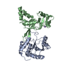
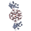
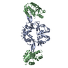
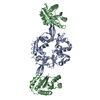
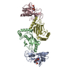
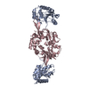
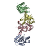
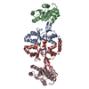
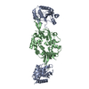
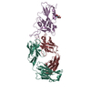
| Title | Crystal structure of PRL-3 phosphatase in complex with the Bateman domain of CNNM3 magnesium transporter | ||||||
 Components Components |
| ||||||
 Keywords Keywords | METAL TRANSPORT / phosphatase / magnesium transporter / protein binding | ||||||
| Function / homology |  Function and homology information Function and homology informationregulation of vascular endothelial growth factor signaling pathway / magnesium ion homeostasis / positive regulation of vascular permeability / endothelial cell migration / Notch signaling pathway / transmembrane transporter activity / monoatomic ion transport / cellular response to leukemia inhibitory factor / protein-tyrosine-phosphatase / protein tyrosine phosphatase activity ...regulation of vascular endothelial growth factor signaling pathway / magnesium ion homeostasis / positive regulation of vascular permeability / endothelial cell migration / Notch signaling pathway / transmembrane transporter activity / monoatomic ion transport / cellular response to leukemia inhibitory factor / protein-tyrosine-phosphatase / protein tyrosine phosphatase activity / early endosome / membrane / nucleus / plasma membrane / cytoplasm Similarity search - Function | ||||||
| Biological species |  Homo sapiens (human) Homo sapiens (human) | ||||||
| Method |  X-RAY DIFFRACTION / SYNCHROTRON / MOLECULAR REPLACEMENT / Resolution: 3.188 Å X-RAY DIFFRACTION / SYNCHROTRON / MOLECULAR REPLACEMENT / Resolution: 3.188 Å | ||||||
 Authors Authors | Kozlov, G. / Zhang, H. / Gehring, K. | ||||||
 Citation Citation | Journal: Sci Rep / Year: 2017 Title: PRL3 phosphatase active site is required for binding the putative magnesium transporter CNNM3. Authors: Zhang, H. / Kozlov, G. / Li, X. / Wu, H. / Gulerez, I. / Gehring, K. | ||||||
| History |
|
- Structure visualization
| Structure viewer | Molecule: MolmilJmol/JSmol |
|---|
- Downloads & links
-Download
| PDBx/mmCIF format | 5tsr.cif.gz | 132.3 KB | Display | PDBx/mmCIF format |
|---|---|---|---|---|
| PDB format | pdb5tsr.ent.gz | 102.2 KB | Display | PDB format |
| PDBx/mmJSON format | 5tsr.json.gz | Tree view | PDBx/mmJSON format | |
| Others |  Other downloads Other downloads |
-Validation report
| Arichive directory | https://data.pdbj.org/pub/pdb/validation_reports/ts/5tsrftp://data.pdbj.org/pub/pdb/validation_reports/ts/5tsr | HTTPS FTP |
|---|
-Related structure data
| Related structure data |  5k23C  5k24C  5k25C  5k22S S: Starting model for refinement C: citing same article ( |
|---|---|
| Similar structure data |
-Links
 PDBj
PDBj


- Assembly
| Deposited unit | 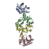
| ||||||||
|---|---|---|---|---|---|---|---|---|---|
| 1 |
| ||||||||
| Unit cell |
|
-Components
| #1: Protein | Mass: 19380.494 Da / Num. of mol.: 2 / Mutation: C104A Source method: isolated from a genetically manipulated source Source: (gene. exp.) Homo sapiens (human) / Gene: PTP4A3, PRL3 / Plasmid: pET15b / Production host:  #2: Protein | Mass: 17634.027 Da / Num. of mol.: 2 Source method: isolated from a genetically manipulated source Source: (gene. exp.) Homo sapiens (human) / Gene: CNNM3, ACDP3 / Plasmid: pDEST15 / Production host: #3: Water | ChemComp-HOH / |  Mass: 18.015 Da / Num. of mol.: 3 / Source method: isolated from a natural source / Formula: H2O Mass: 18.015 Da / Num. of mol.: 3 / Source method: isolated from a natural source / Formula: H2O |
|---|
-Experimental details
-Experiment
| Experiment | Method: X-RAY DIFFRACTION / Number of used crystals: 1 |
|---|
- Sample preparation
| Crystal | Density Matthews: 3.34 Å3/Da / Density % sol: 63.17 % |
|---|---|
| Crystal grow | Temperature: 293 K / Method: vapor diffusion, hanging drop Details: 0.1 M HEPES pH 7.0, 0.064 M Tri-Na Citrate, 15% PEG 5000 MME |
-Data collection
| Diffraction | Mean temperature: 100 K |
|---|---|
| Diffraction source | Source: SYNCHROTRON / Site: CHESS  / Beamline: F1 / Wavelength: 0.977 Å / Beamline: F1 / Wavelength: 0.977 Å |
| Detector | Type: DECTRIS PILATUS3 S 6M / Detector: PIXEL / Date: Jun 28, 2016 |
| Radiation | Protocol: SINGLE WAVELENGTH / Monochromatic (M) / Laue (L): M / Scattering type: x-ray |
| Radiation wavelength | Wavelength: 0.977 Å / Relative weight: 1 |
| Reflection | Resolution: 3.188→50 Å / Num. obs: 15609 / % possible obs: 96.6 % / Redundancy: 3.3 % / Rsym value: 0.106 / Net I/σ(I): 10.5 |
| Reflection shell | Resolution: 3.188→3.26 Å / Redundancy: 3 % / Rmerge(I) obs: 0.534 / Mean I/σ(I) obs: 1.5 / % possible all: 99.6 |
- Processing
| Software |
| |||||||||||||||||||||||||||||||||||||||||||||||||
|---|---|---|---|---|---|---|---|---|---|---|---|---|---|---|---|---|---|---|---|---|---|---|---|---|---|---|---|---|---|---|---|---|---|---|---|---|---|---|---|---|---|---|---|---|---|---|---|---|---|---|
| Refinement | Method to determine structure: MOLECULAR REPLACEMENT Starting model: 5K22 Resolution: 3.188→27.738 Å / SU ML: 0.57 / Cross valid method: THROUGHOUT / σ(F): 1.4 / Phase error: 32.86
| |||||||||||||||||||||||||||||||||||||||||||||||||
| Solvent computation | Shrinkage radii: 0.9 Å / VDW probe radii: 1.11 Å | |||||||||||||||||||||||||||||||||||||||||||||||||
| Refinement step | Cycle: LAST / Resolution: 3.188→27.738 Å
| |||||||||||||||||||||||||||||||||||||||||||||||||
| Refine LS restraints |
| |||||||||||||||||||||||||||||||||||||||||||||||||
| LS refinement shell |
|
