 Movie
Movie Controller

[English] 日本語
 Yorodumi
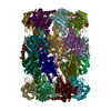
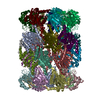
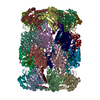
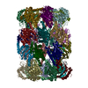
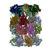
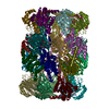
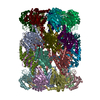
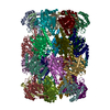
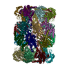
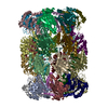
- PDB-5trr: Structure of Mycobacterium tuberculosis proteasome in complex wit... -
+ Open data

- Basic information
| Entry | Database: PDB / ID: 5trr | ||||||
|---|---|---|---|---|---|---|---|
| Title | Structure of Mycobacterium tuberculosis proteasome in complex with N,C-capped dipeptide PKS2169 | ||||||
 Components Components |
| ||||||
 Keywords Keywords | HYDROLASE/HYDROLASE inhibitor / N / C-capped dipeptides / Mycobacterium tuberculosis / proteasome inhibitors / HYDROLASE-HYDROLASE inhibitor complex | ||||||
| Function / homology |  Function and homology information Function and homology informationsymbiont-mediated perturbation of host defenses / zymogen binding / proteasome endopeptidase complex / proteasome core complex, beta-subunit complex / threonine-type endopeptidase activity / proteasome core complex, alpha-subunit complex / peptidoglycan-based cell wall / : / proteasomal protein catabolic process / modification-dependent protein catabolic process ...symbiont-mediated perturbation of host defenses / zymogen binding / proteasome endopeptidase complex / proteasome core complex, beta-subunit complex / threonine-type endopeptidase activity / proteasome core complex, alpha-subunit complex / peptidoglycan-based cell wall / : / proteasomal protein catabolic process / modification-dependent protein catabolic process / proteasome-mediated ubiquitin-dependent protein catabolic process / extracellular region / plasma membrane / cytoplasm Similarity search - Function | ||||||
| Biological species |   Mycobacterium tuberculosis (bacteria) Mycobacterium tuberculosis (bacteria) | ||||||
| Method |  X-RAY DIFFRACTION / SYNCHROTRON / MOLECULAR REPLACEMENT / Resolution: 3.103 Å X-RAY DIFFRACTION / SYNCHROTRON / MOLECULAR REPLACEMENT / Resolution: 3.103 Å | ||||||
 Authors Authors | Hsu, H.-C. / Fan, H. / Singh, P.K. / Wang, R. / Sukenick, G. / Nathan, C. / Lin, G. / Li, H. | ||||||
| Funding support |  United States, 1items United States, 1items
| ||||||
 Citation Citation | Journal: Biochemistry / Year: 2017 Title: Structural Basis for the Species-Selective Binding of N,C-Capped Dipeptides to the Mycobacterium tuberculosis Proteasome. Authors: Hsu, H.C. / Singh, P.K. / Fan, H. / Wang, R. / Sukenick, G. / Nathan, C. / Lin, G. / Li, H. | ||||||
| History |
|
- Structure visualization
| Structure viewer | Molecule: MolmilJmol/JSmol |
|---|
- Downloads & links
-Download
| PDBx/mmCIF format | 5trr.cif.gz | 1.1 MB | Display | PDBx/mmCIF format |
|---|---|---|---|---|
| PDB format | pdb5trr.ent.gz | 936.2 KB | Display | PDB format |
| PDBx/mmJSON format | 5trr.json.gz | Tree view | PDBx/mmJSON format | |
| Others |  Other downloads Other downloads |
-Validation report
| Arichive directory | https://data.pdbj.org/pub/pdb/validation_reports/tr/5trrftp://data.pdbj.org/pub/pdb/validation_reports/tr/5trr | HTTPS FTP |
|---|
-Related structure data
| Related structure data | 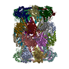 5thoC 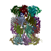 5trgC 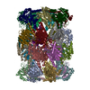 5trsC  5tryC 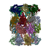 5ts0C  3hfaS C: citing same article ( S: Starting model for refinement |
|---|---|
| Similar structure data |


-Links
 PDBj
PDBj


- Assembly
| Deposited unit | 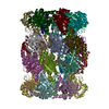
| ||||||||||
|---|---|---|---|---|---|---|---|---|---|---|---|
| 1 |
| ||||||||||
| Unit cell |
|
-Components
| #1: Protein | Mass: 25971.975 Da / Num. of mol.: 14 / Fragment: UNP residues 10-248 Source method: isolated from a genetically manipulated source Source: (gene. exp.) Mycobacterium tuberculosis (bacteria) / Gene: prcA, MRA_2124 / Production host: References: UniProt: A5U4D5, UniProt: P9WHU1*PLUS, proteasome endopeptidase complex #2: Protein | Mass: 25274.264 Da / Num. of mol.: 14 / Fragment: UNP residues 58-291 Source method: isolated from a genetically manipulated source Source: (gene. exp.) Mycobacterium tuberculosis (bacteria) / Gene: prcB, MRA_2125 / Production host: References: UniProt: A5U4D6, UniProt: P9WHT9*PLUS, proteasome endopeptidase complex #3: Chemical | ChemComp-7HY /   Type: Non-polymer / Class: Inhibitor / Mass: 530.658 Da / Num. of mol.: 14 / Source method: obtained synthetically / Formula: C31H38N4O4 Type: Non-polymer / Class: Inhibitor / Mass: 530.658 Da / Num. of mol.: 14 / Source method: obtained synthetically / Formula: C31H38N4O4References: N,N-diethyl-N~2~-(3-phenylpropanoyl)-L-asparaginyl-N-[(naphthalen-1-yl)methyl]-L-alaninamide #4: Water | ChemComp-HOH / |  Mass: 18.015 Da / Num. of mol.: 62 / Source method: isolated from a natural source / Formula: H2O Mass: 18.015 Da / Num. of mol.: 62 / Source method: isolated from a natural source / Formula: H2O |
|---|
-Experimental details
-Experiment
| Experiment | Method: X-RAY DIFFRACTION / Number of used crystals: 1 |
|---|
- Sample preparation
| Crystal | Density Matthews: 2.67 Å3/Da / Density % sol: 53.89 % |
|---|---|
| Crystal grow | Temperature: 277 K / Method: vapor diffusion, hanging drop / pH: 6.2 / Details: 60 mM sodium citrate (pH 6.2) and 14% PEG-3350 |
-Data collection
| Diffraction | Mean temperature: 100 K |
|---|---|
| Diffraction source | Source: SYNCHROTRON / Site: NSLS / Beamline: X25 / Wavelength: 1.1 Å |
| Detector | Type: DECTRIS PILATUS 6M / Detector: PIXEL / Date: Sep 15, 2014 |
| Radiation | Protocol: SINGLE WAVELENGTH / Monochromatic (M) / Laue (L): M / Scattering type: x-ray |
| Radiation wavelength | Wavelength: 1.1 Å / Relative weight: 1 |
| Reflection | Resolution: 3.1→47.2473073393 Å / Num. obs: 131508 / % possible obs: 99.5 % / Redundancy: 3.5 % / Biso Wilson estimate: 73.5256458866 Å2 / CC1/2: 0.98 / Rmerge(I) obs: 0.106 / Net I/σ(I): 7.6 |
| Reflection shell | Resolution: 3.1→3.2 Å / Redundancy: 3.2 % / Rmerge(I) obs: 0.483 / Mean I/σ(I) obs: 2.5 / CC1/2: 0.775 / % possible all: 95.1 |
- Processing
| Software |
| |||||||||||||||||||||||||||||||||||||||||||||||||||||||||||||||||||||||||||||||||||||||||||||||||||||||||||||||||||||||||||||||||||||||||||||||||||||||||||||||||||||||||||||||||||||||||||||||||||||||||||||||||||||||||
|---|---|---|---|---|---|---|---|---|---|---|---|---|---|---|---|---|---|---|---|---|---|---|---|---|---|---|---|---|---|---|---|---|---|---|---|---|---|---|---|---|---|---|---|---|---|---|---|---|---|---|---|---|---|---|---|---|---|---|---|---|---|---|---|---|---|---|---|---|---|---|---|---|---|---|---|---|---|---|---|---|---|---|---|---|---|---|---|---|---|---|---|---|---|---|---|---|---|---|---|---|---|---|---|---|---|---|---|---|---|---|---|---|---|---|---|---|---|---|---|---|---|---|---|---|---|---|---|---|---|---|---|---|---|---|---|---|---|---|---|---|---|---|---|---|---|---|---|---|---|---|---|---|---|---|---|---|---|---|---|---|---|---|---|---|---|---|---|---|---|---|---|---|---|---|---|---|---|---|---|---|---|---|---|---|---|---|---|---|---|---|---|---|---|---|---|---|---|---|---|---|---|---|---|---|---|---|---|---|---|---|---|---|---|---|---|---|---|---|
| Refinement | Method to determine structure: MOLECULAR REPLACEMENT Starting model: 3HFA Resolution: 3.103→47.247 Å / SU ML: 0.32 / Cross valid method: FREE R-VALUE / σ(F): 1.33 / Phase error: 21.07 / Stereochemistry target values: ML
| |||||||||||||||||||||||||||||||||||||||||||||||||||||||||||||||||||||||||||||||||||||||||||||||||||||||||||||||||||||||||||||||||||||||||||||||||||||||||||||||||||||||||||||||||||||||||||||||||||||||||||||||||||||||||
| Solvent computation | Shrinkage radii: 0.9 Å / VDW probe radii: 1.11 Å / Solvent model: FLAT BULK SOLVENT MODEL | |||||||||||||||||||||||||||||||||||||||||||||||||||||||||||||||||||||||||||||||||||||||||||||||||||||||||||||||||||||||||||||||||||||||||||||||||||||||||||||||||||||||||||||||||||||||||||||||||||||||||||||||||||||||||
| Refinement step | Cycle: LAST / Resolution: 3.103→47.247 Å
| |||||||||||||||||||||||||||||||||||||||||||||||||||||||||||||||||||||||||||||||||||||||||||||||||||||||||||||||||||||||||||||||||||||||||||||||||||||||||||||||||||||||||||||||||||||||||||||||||||||||||||||||||||||||||
| Refine LS restraints |
| |||||||||||||||||||||||||||||||||||||||||||||||||||||||||||||||||||||||||||||||||||||||||||||||||||||||||||||||||||||||||||||||||||||||||||||||||||||||||||||||||||||||||||||||||||||||||||||||||||||||||||||||||||||||||
| LS refinement shell |
|
