- PDB-5tro: 1.8 Angstrom Resolution Crystal Structure of Dimerization and Tra... -
+
Open data
ID or keywords:
Loading...
-
Basic information
Entry
Database: PDB / ID: 5tro
Title
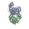
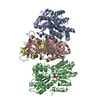
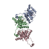
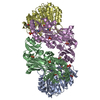
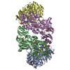
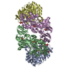
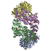
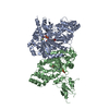
1.8 Angstrom Resolution Crystal Structure of Dimerization and Transpeptidase domains (residues 39-608) of Penicillin-Binding Protein 1 from Staphylococcus aureus.
Components
Penicillin-binding protein 1
Keywords
HYDROLASE / Structural Genomics / Center for Structural Genomics of Infectious Diseases / CSGID / Dimerization Domain / Transpeptidase Domain / Penicillin-Binding Protein 1
Type: MARMOSAIC 300 mm CCD / Detector: CCD / Date: Oct 20, 2016 / Details: C(111)
Radiation
Monochromator: beryllium lenses / Protocol: SINGLE WAVELENGTH / Monochromatic (M) / Laue (L): M / Scattering type: x-ray
Radiation wavelength
Wavelength: 0.97872 Å / Relative weight: 1
Reflection
Resolution: 1.8→30 Å / Num. obs: 98204 / % possible obs: 99.7 % / Observed criterion σ(I): -3 / Redundancy: 5.4 % / Biso Wilson estimate: 30.3 Å2 / Rmerge(I) obs: 0.088 / Rsym value: 0.088 / Net I/σ(I): 26.8
Reflection shell
Resolution: 1.8→1.83 Å / Redundancy: 4.9 % / Rmerge(I) obs: 0.749 / Mean I/σ(I) obs: 2.3 / CC1/2: 0.793 / % possible all: 99.3
-
Processing
Software
Name
Version
Classification
REFMAC
5.8.0155
refinement
HKL-3000
datareduction
HKL-3000
datascaling
PHENIX
phasing
Refinement
Method to determine structure: SAD / Resolution: 1.8→29.59 Å / Cor.coef. Fo:Fc: 0.966 / Cor.coef. Fo:Fc free: 0.955 / SU B: 6.435 / SU ML: 0.098 / Cross valid method: THROUGHOUT / ESU R: 0.127 / ESU R Free: 0.118 / Details: HYDROGENS HAVE BEEN ADDED IN THE RIDING POSITIONS
Rfactor
Num. reflection
% reflection
Selection details
Rfree
0.20946
4950
5 %
RANDOM
Rwork
0.17935
-
-
-
obs
0.18088
93238
99.22 %
-
Solvent computation
Ion probe radii: 0.8 Å / Shrinkage radii: 0.8 Å / VDW probe radii: 1.2 Å
 Movie
Movie Controller

 Yorodumi
 Open data
 Basic information
 Components
 Keywords
 Function and homology information
 Staphylococcus aureus (bacteria)
 X-RAY DIFFRACTION /
  Authors
 Citation
 Structure visualization
 Downloads & links
 Other downloads
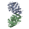
 PDBj
PDBj Assembly
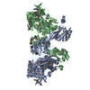

 Mass: 35.453 Da / Num. of mol.: 2 / Source method: obtained synthetically / Formula: Cl
 Mass: 18.015 Da / Num. of mol.: 615 / Source method: isolated from a natural source / Formula: H2O
 Sample preparation
 / Beamline: 21-ID-F / Wavelength: 0.97872 Å
 Processing